Chimera of alcohol dehydrogenase by exchange of the cofactor binding domain res 153-294 of T. brockii ADH by E. histolytica ADH
From Proteopedia
| |||||||
| Chimera of alcohol dehydrogenase complex with etylene glycol, nitrate, cacodylate, O2, imidazole, Na+ and Zn+2 ions 3fpc | |||||||
|---|---|---|---|---|---|---|---|
| Ligands: | , , , , , , | ||||||
| Gene: | ADH1, ADH1 (ENTHI) | ||||||
| Activity: | Alcohol dehydrogenase (NADP(+)), with EC number 1.1.1.2 | ||||||
| Related: | 3fpl | ||||||
| |||||||
| |||||||
| Resources: | FirstGlance, OCA, RCSB, PDBsum | ||||||
| Coordinates: | save as pdb, mmCIF, xml | ||||||
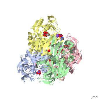


Chimera Χ23(TET) (3fpc)
The cofactor-binding domains (residues 153-295) of the alcohol dehydrogenases from the thermophile Thermoanaerobacter brockii (TbADH), the mesophilic bacterium Clostridium beijerinckii (CbADH), and the protozoan parasite Entamoeba histolytica (EhADH1) have been exchanged. Three chimeras have been constructed. In the first chimera, the cofactor-binding domain of thermophilic TbADH was replaced with the cofactor-binding domain of its mesophilic counterpart CbADH [chimera Chi21((TCT))]. This domain exchange significantly destabilized the parent thermophilic enzyme (DeltaT(1/2) = -18 degrees C). The reverse exchange in CbADH [chimera Chi22((CTC))], however, had little effect on the thermal stability of the parent mesophilic protein. Furthermore, substituting the cofactor-binding domain of TbADH with the homologous domain of EhADH1 [chimera Chi23((TET))] substantially reduced the thermal stability of the thermophilic ADH (DeltaT(1/2) = -51 degrees C) and impeded the oligomerization of the enzyme. All three chimeric proteins and one of their site-directed mutants were crystallized, and their three-dimensional (3D) structures were determined. Comparison of the 3D structures of the chimeras and the chimeric mutant with the structures of their parent ADHs showed no significant changes to their Calpha chains, suggesting that the difference in the thermal stability of the three parent ADHs and their chimeric mutants could be due to a limited number of substitutions located at strategic positions, mainly at the oligomerization interfaces. Indeed, stabilization of the chimeras was achieved, to a significant extent, either by introduction of a proline residue at a strategic position in the major horse liver ADH-type dimerization interface (DeltaT(1/2) = 35 degrees C) or by introduction of intersubunit electrostatic interactions (DeltaT(1/2) = 6 degrees C).
Biochemical and Structural Properties of Chimeras Constructed by Exchange of Cofactor-Binding Domains in Alcohol Dehydrogenases from Thermophilic and Mesophilic Microorganisms., Goihberg E, Peretz M, Tel-Or S, Dym O, Shimon L, Frolow F, Burstein Y, Biochemistry. 2010 Feb 9. PMID:20102159
From MEDLINE®/PubMed®, a database of the U.S. National Library of Medicine.
The NADP+-dependent alcohol dehydrogenases (EC 1.1.1.2) from the thermophile Thermoanaerobacter brockii (TbADH), the mesophilic bacterium Clostridium beijerinckii (CbADH), and the protozoan parasite Entamoeba histolytica (EhADH1) are [1] (monomers are colored in different colors) secondary alcohol dehydrogenases. Each of these alcohol dehydrogenases consists of two domains: the (residues 154−294 for TbADH) and the (residues 1−153 and 295−351 for TbADH; contains Zn2+ at the active site) separated by a deep cleft. Although, all these three ADHs revealed a high degree of sequence conservation (62-75% identity), them significantly differ in thermostability. The cofactor-binding domains (residues 153−295) of TbADH, CbADH, and EhADH1 were mutually and 3 corresponding chimeras were constructed.
The exchange of the cofactor-binding domain of TbADH with the homologous domain of EhADH1 (chimera Χ23(TET), 3fpc) substantially reduced the thermal stability of the thermophilic ADH (ΔT1/2 = −51 °C) and interfered the oligomerization of the enzyme.
The of overall Cα backbone of all these chimeras (rmsd 0.45-0.65 Å) with those of the parent enzymes, did not reveal significant structural changes. So, the differences in the thermal stability of the chimeras and the parent enzymes could be caused by relatively small specific changes located at the important points of the NADP+-dependent alcohol dehydrogenases. For example see Cα superposition for the X23(TET) chimera (red) (3fpc) and its parent ADHs (TbADH, colored blue (1ped), and EhADH1, colored lime (1y9a). The RMSDs of the TbADH−EhADH1, TbADH−Χ23(TET), and EhADH1−Χ23(TET) were 0.68, 0.56, and 0.48 Å, respectively.
</StructureSection>
Reference
- Goihberg E, Peretz M, Tel-Or S, Dym O, Shimon L, Frolow F, Burstein Y. Biochemical and Structural Properties of Chimeras Constructed by Exchange of Cofactor-Binding Domains in Alcohol Dehydrogenases from Thermophilic and Mesophilic Microorganisms. Biochemistry. 2010 Feb 9. PMID:20102159 doi:10.1021/bi901730x
- (See also Tetrameric alcohol dehydrogenases)
- (See also Chimeres of alcohol dehydrogenases)
Proteopedia Page Contributors and Editors (what is this?)
Categories: Thermoanaerobacter brockii, entamoeba histolytica | Burstein, Y. | Felix, F. | Goihberg, E. | Shimon, L. | Bacterial alcohol dehydrogenase | Chimera | Cytoplasm | Domain exchange | Metal-binding | Nadp | Oxidoreductase | Zinc | ISPC, Israel Structural Proteomics Center. | Peretz, M. | Tel-Or, S. | ISPC | Israel Structural Proteomics Center | Structural genomic
