Image:Anthrax-toxin.gif
From Proteopedia
No higher resolution available.
Anthrax-toxin.gif (640 × 361 pixel, file size: 100 KB, MIME type: image/gif)
Contents |
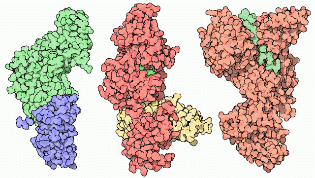
3D Printed Physical Model of the Anthrax Toxin Protein
Shown below are 3D printed physical models of the Anthrax Toxin Protein, based on the structure 1acc.pdb. The full septamer model is shown in spacefill format and is colored by domain, with a single monomer shown in white. The alpha carbon backbone model is similarly colored by chain, with additional key side chains included.


The MSOE Center for BioMolecular Modeling

The MSOE Center for BioMolecular Modeling uses 3D printing technology to create physical models of protein and molecular structures, making the invisible molecular world more tangible and comprehensible. To view more protein structure models, visit our Model Gallery.
Summary

Licensing
{{subst:No license from license selector|Don't know}}
File history
Click on a date/time to view the file as it appeared at that time.
| Date/Time | User | Dimensions | File size | Comment | |
|---|---|---|---|---|---|
| (current) | 13:55, 6 January 2013 | Charlotte Kern (Talk | contribs) | 640×361 | 100 KB | http://www.rcsb.org/pdb/education_discussion/molecule_of_the_month/images/anthrax-toxin.gif |
- Edit this file using an external application
See the setup instructions for more information.
Links
The following pages link to this file:
