Image:Interaction 1 between Bmi1.Ring1b and UbcH5c.png
From Proteopedia
No higher resolution available.
Interaction_1_between_Bmi1.Ring1b_and_UbcH5c.png (484 × 323 pixel, file size: 170 KB, MIME type: image/png)
File history
Click on a date/time to view the file as it appeared at that time.
| Date/Time | User | Dimensions | File size | Comment | |
|---|---|---|---|---|---|
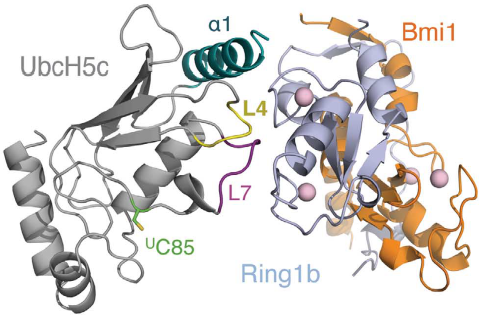
| (current) | 12:30, 17 June 2018 | Ricardo Alberto Chiong Zevallos (Talk | contribs) | 484×323 | 170 KB | Structure of the Bmi1/Ring1b–UbcH5c complex. (A) (Left) Ribbon representation of the overall complex architecture, with UbcH5c in grey, Ring1b(1116) in light blue, and Bmi1(1109) in orange. Zn2þ ions are shown in pink. The three Ring1b-binding regions |
| 23:11, 15 June 2018 | Ricardo Alberto Chiong Zevallos (Talk | contribs) | 484×323 | 170 KB |
- Edit this file using an external application
See the setup instructions for more information.
Links
The following pages link to this file:
