Acid phosphatase
From Proteopedia
FunctionAcid phosphatase (ACP, EC number 3.1.3.2) is an enzyme which removes phosphate from other molecules during digestion. It catalyzes the conversion of orthophosphoric monoester and H2O to alcohol and phosphoric acid. The enzyme is most effective in acidic environment, hence its name.[1]
ACP contains 3 classes:
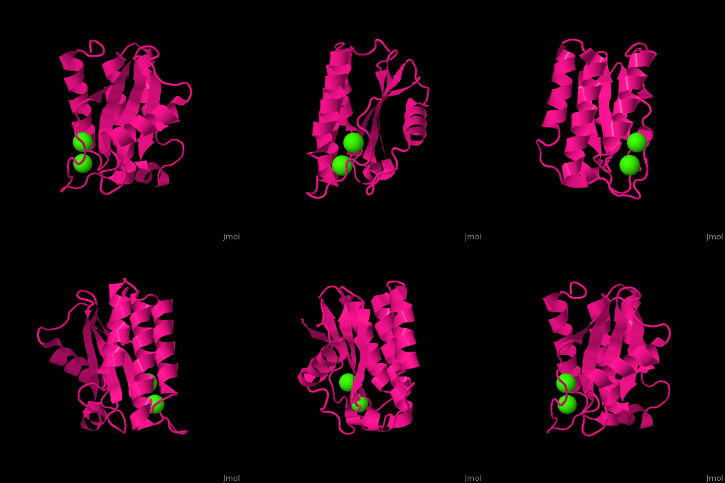
In plants, the TPM domain-containing proteins TLP18.3 and Psb32 that have been implicated in the photosystem II (PSII) repair cycle. It may be involved in the regulation of synthesis/degradation of the D1 protein of the PSII core and in the assembly of PSII monomers into dimers in the grana stacks.[8] In the model nematode C. elegans, the MOLO-1 protein is an auxiliary subunit that positively modulates the gating of levamisole-sensitive acetylcholine receptors.[9] 2nd Ca2+ coordination site in Antarctic bacterium protein BA42 (PDB code 4oa3).[10] DiseasePSAP is found in increased amounts in patients who have prostate cancer. RelevancePSAP was used as a prostate cancer marker before the develpement of prostate specific antigen (PSA) as one. Structural HighlightsHg2+ cation acting as intermolecular bridge in E. coli acid phosphatase.[11] 3D Structures of acid phosphataseAcid phosphatase 3D structures
|
| |||||||||||
References
- ↑ Bull H, Murray PG, Thomas D, Fraser AM, Nelson PN. Acid phosphatases. Mol Pathol. 2002 Apr;55(2):65-72. PMID:11950951
- ↑ Hassan MI, Aijaz A, Ahmad F. Structural and functional analysis of human prostatic acid phosphatase. Expert Rev Anticancer Ther. 2010 Jul;10(7):1055-68. PMID:20645695 doi:10.1586/era.10.46
- ↑ Bhadouria J, Giri J. Purple acid phosphatases: roles in phosphate utilization and new emerging functions. Plant Cell Rep. 2022 Jan;41(1):33-51. PMID:34402946 doi:10.1007/s00299-021-02773-7
- ↑ Rigden DJ. The histidine phosphatase superfamily: structure and function. Biochem J. 2008 Jan 15;409(2):333-48. PMID:18092946 doi:10.1042/BJ20071097
- ↑ Reddy VS, Rao DK, Rajasekharan R. Functional characterization of lysophosphatidic acid phosphatase from Arabidopsis thaliana. Biochim Biophys Acta. 2010 Apr;1801(4):455-61. PMID:20045079 doi:10.1016/j.bbalip.2009.12.005
- ↑ Wu HY, Liu MS, Lin TP, Cheng YS. Structural and Functional Assays of AtTLP18.3 Identify Its Novel Acid Phosphatase Activity in Thylakoid Lumen. Plant Physiol. 2011 Sep 9. PMID:21908686 doi:10.1104/pp.111.184739
- ↑ Eletsky A, Acton TB, Xiao R, Everett JK, Montelione GT, Szyperski T. Solution NMR structures reveal a distinct architecture and provide first structures for protein domain family PF04536. J Struct Funct Genomics. 2012 Mar;13(1):9-14. doi: 10.1007/s10969-011-9122-2., Epub 2011 Dec 24. PMID:22198206 doi:http://dx.doi.org/10.1007/s10969-011-9122-2
- ↑ Sirpio S, Allahverdiyeva Y, Suorsa M, Paakkarinen V, Vainonen J, Battchikova N, Aro EM. TLP18.3, a novel thylakoid lumen protein regulating photosystem II repair cycle. Biochem J. 2007 Sep 15;406(3):415-25. PMID:17576201 doi:http://dx.doi.org/10.1042/BJ20070460
- ↑ Boulin T, Rapti G, Briseno-Roa L, Stigloher C, Richmond JE, Paoletti P, Bessereau JL. Positive modulation of a Cys-loop acetylcholine receptor by an auxiliary transmembrane subunit. Nat Neurosci. 2012 Oct;15(10):1374-81. doi: 10.1038/nn.3197. Epub 2012 Aug 26. PMID:22922783 doi:http://dx.doi.org/10.1038/nn.3197
- ↑ Aran M, Smal C, Pellizza L, Gallo M, Otero LH, Klinke S, Goldbaum FA, Ithurralde ER, Bercovich A, Mac Cormack WP, Turjanski AG, Cicero DO. Solution and crystal structure of BA42, a protein from the Antarctic bacterium Bizionia argentinensis comprised of a stand-alone TPM domain. Proteins. 2014 Aug 13. doi: 10.1002/prot.24667. PMID:25116514 doi:http://dx.doi.org/10.1002/prot.24667
- ↑ Lim D, Golovan S, Forsberg CW, Jia Z. Crystal structures of Escherichia coli phytase and its complex with phytate. Nat Struct Biol. 2000 Feb;7(2):108-13. PMID:10655611 doi:http://dx.doi.org/10.1038/72371

