File:GLUT1 Binding Mechanism.png
Original file (1,700 × 775 pixels, file size: 541 KB, MIME type: image/png)
Proteopedia Page Contributors and Editors (what is this?)
Summary
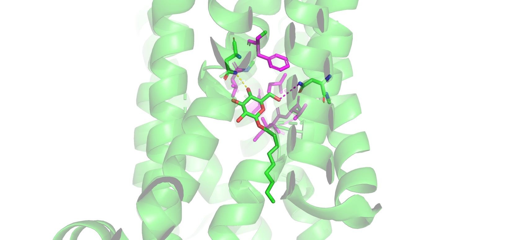
Proposed glucose-binding mechanism in the crystal structure 4pyp rendered in PyMol. The proposed hydrophobic pocket residues of Gly27, Thr30, Ile164, Val165, Ile168, and Phe291 are colored magenta. Both Asn288 and Asn317 show that there may also be transient hydrogen bonding in glucose binding to GLUT1.
Licensing

I, the copyright holder of this work, hereby release it into the public domain. This applies worldwide.
In case this is not legally possible,
I grant any entity the right to use this work for any purpose, without any conditions, unless such conditions are required by law.
Proteopedia Page Contributors and Editors (what is this?)
File history
Click on a date/time to view the file as it appeared at that time.
| Date/Time | Thumbnail | Dimensions | User | Comment | |
|---|---|---|---|---|---|
| current | 22:00, 2 May 2022 | | 1,700 × 775 (541 KB) | Adam Kingsley (talk | contribs) | Proposed glucose-binding mechanism in the crystal structure 4pyp rendered in PyMol. The proposed hydrophobic pocket residues of Gly27, Thr30, Ile164, Val165, Ile168, and Phe291 are colored magenta. Both Asn288 and Asn317 show that there may also be tr |
You cannot overwrite this file.
File usage
There are no pages that use this file.


