File:Gabab.xu2014.png
From Proteopedia
Jump to navigationJump to search
No higher resolution available.
Gabab.xu2014.png (326 × 308 pixels, file size: 50 KB, MIME type: image/png)
Proteopedia Page Contributors and Editors (what is this?)
Summary
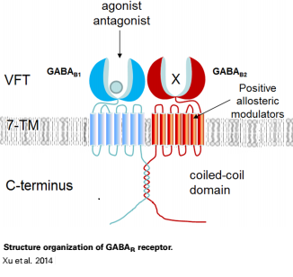
Structure organization of GABAB receptor. GABAB receptor forms heterodimer composed by GABAB1 and GABAB2. GABAB1 is responsible for ligand binding in N-terminal VFT domain, whereas the VFT of GABAB2 fails to bind any known ligand (Xu et al.2014).
Licensing
{{subst:Non-commercial from license selector}}
Proteopedia Page Contributors and Editors (what is this?)
File history
Click on a date/time to view the file as it appeared at that time.
| Date/Time | Thumbnail | Dimensions | User | Comment | |
|---|---|---|---|---|---|
| current | 06:55, 18 May 2015 | | 326 × 308 (50 KB) | Rana Saad (talk | contribs) | Structure organization of GABAB receptor. GABAB receptor forms heterodimer composed by GABAB1 and GABAB2. GABAB1 is responsible for ligand binding in N-terminal VFT domain, whereas the VFT of GABAB2 fails to bind any known ligand (Xu et al.2014). |
You cannot overwrite this file.
File usage
The following page uses this file:


