File:Glycogen breakdown 3.png
From Proteopedia
Jump to navigationJump to search
Size of this preview: 692 × 600 pixels. Other resolution: 1,125 × 975 pixels.
Original file (1,125 × 975 pixels, file size: 18 KB, MIME type: image/png)
Proteopedia Page Contributors and Editors (what is this?)
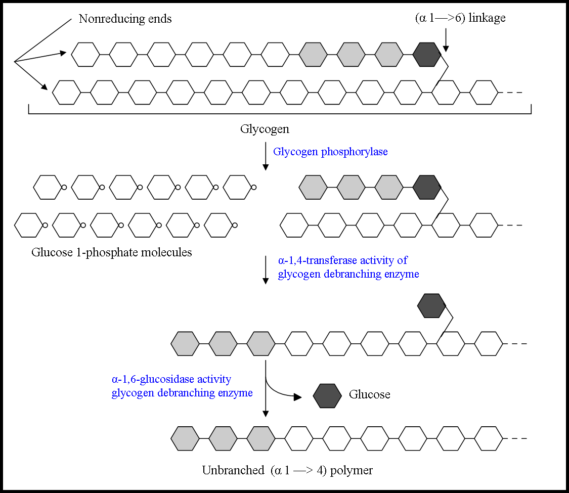
Figure 1. Diagram illustrating the breakdown of glycogen near an (α1→6) branch point, and the steps where the α-1,6-glycosidase and the α-1,4-transferase activity of the glycogen debranching enzyme takes place. Diagram adapted from Principles of Biochemistry 5th ed.
Proteopedia Page Contributors and Editors (what is this?)
File history
Click on a date/time to view the file as it appeared at that time.
| Date/Time | Thumbnail | Dimensions | User | Comment | |
|---|---|---|---|---|---|
| current | 22:57, 1 April 2011 | | 1,125 × 975 (18 KB) | Nikolina Nikolic (talk | contribs) | Figure 1. Diagram illustrating the breakdown of glycogen near an (α1→6) branch point, and the steps where the α-1,6-glycosidase and the α-1,4-transferase activity of the glycogen debranching enzyme takes place. Diagram adapted from Principles of Bioc |
| 22:56, 1 April 2011 |  | 1,125 × 975 (18 KB) | Nikolina Nikolic (talk | contribs) | Figure 1. Diagram illustrating the breakdown of glycogen near an (α1→6) branch point, and the steps where the α-1,6-glycosidase and the α-1,4-transferase activity of the glycogen debranching enzyme takes place. Diagram adapted from Principles of Bioc |
You cannot overwrite this file.
File usage
The following page uses this file:


