File:Tertiary Structure.gif
From Proteopedia
Jump to navigationJump to search
Size of this preview: 458 × 599 pixels. Other resolution: 500 × 654 pixels.
Original file (500 × 654 pixels, file size: 78 KB, MIME type: image/gif)
Proteopedia Page Contributors and Editors (what is this?)
Summary
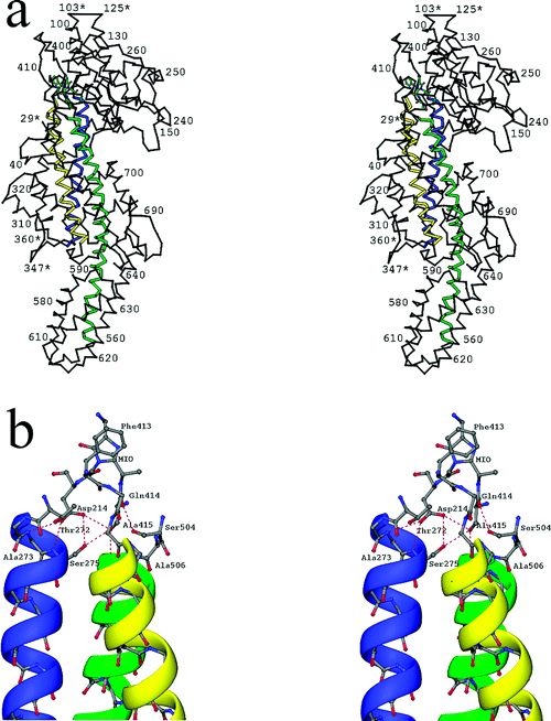
Tertiary structure of PAL. The three central core helices, leading to the active site, are colored blue, green, and yellow for emphasis. (a) Stereoview of the PAL monomer with residue numbering. Breaks in the polypeptide chain are indicated by asterisks. (b) Close-up stereoview of MIO and Phe413 interactions with the three central helices, polarized with their N termini directed toward the active site. Hydrogen bonds are indicated by dashed lines.
Licensing
{{subst:Non-commercial from license selector}}
Calabrese et al [1]
Proteopedia Page Contributors and Editors (what is this?)
File history
Click on a date/time to view the file as it appeared at that time.
| Date/Time | Thumbnail | Dimensions | User | Comment | |
|---|---|---|---|---|---|
| current | 13:37, 6 December 2013 | | 500 × 654 (78 KB) | Bryan Toton (talk | contribs) | Tertiary structure of PAL. The three central core helices, leading to the active site, are colored blue, green, and yellow for emphasis. (a) Stereoview of the PAL monomer with residue numbering. Breaks in the polypeptide chain are indicated by asterisks. |
You cannot overwrite this file.
File usage
The following page uses this file:


