File:3h90sections.PNG: Difference between revisions
From Proteopedia
Jump to navigationJump to search
No edit summary |
No edit summary |
||
| Line 1: | Line 1: | ||
The distribution of YiiP through the membrane is shown. The CTD is shown highlighted in the yellow | The distribution of YiiP through the membrane is shown. The CTD is shown highlighted in the yellow to show that it is in the cytoplasm and the TMD is highlighted with blue to show that it sits in the membrane. | ||
Latest revision as of 18:39, 21 April 2017
The distribution of YiiP through the membrane is shown. The CTD is shown highlighted in the yellow to show that it is in the cytoplasm and the TMD is highlighted with blue to show that it sits in the membrane.
Proteopedia Page Contributors and Editors (what is this?)
File history
Click on a date/time to view the file as it appeared at that time.
| Date/Time | Thumbnail | Dimensions | User | Comment | |
|---|---|---|---|---|---|
| current | 18:05, 21 April 2017 | 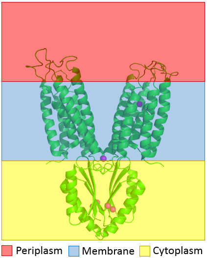 | 423 × 527 (101 KB) | Kyle Colston (talk | contribs) | The distribution of YiiP through the membrane is shown. The CTD is shown in highlighted in the yellow bottom to show that it is in the cytoplasm and the TMD is highlighted with blue to show that it sits in membrane. |
You cannot overwrite this file.
File usage
The following page uses this file:


