You may include any references to papers as in: the use of JSmol in Proteopedia [1] or to the article describing Jmol [2] to the rescue.
Functions and Biological Relevance
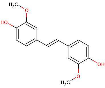
Lignostilbene-α,ß-dioxygenase A (LsdA) from the bacterium Sphingomonas paucimobilis TMY1009 is a nonheme iron oxygenase that catalyzes the cleavage via oxygenolytic fission of lignostilbene, a compound arising in lignin transformation, to two vanillin molecules (see images below). Lignin is a common component of biomass from industry that scientists are interested in finding ways to break down to simpler and useful materials such as biofuels and commodity chemicals. Though natural occurring lignostilbenoids are rare, they are a very comm on byproduct of industry due to condensation reactions. Other lignin-based stilbenes are thought to be produces from bacterial catabolic processing of diaryl propane and phenylcoumarane [3].
Lignostilbene
Vanillin

Broader Implications
Though lignin is the second most abundant fraction byproduct of lignocellulose, it is currently mostly just combusted for steam and electricity generation. It is rarely valorized into other products like a dispersant for cement and gypsum. One major difficulty in converting and valorizing lignin-based byproducts it their highly variable composition. Both the substrate in which they are located, and the means of extraction affect their end composition and properties. There is much research into finding strong methods of breakdown of lignostilbenoid molecules.
Structural highlights and structure-function relationships
LsdA appears, when crystallized, as two LsdA protomers in one asymmetric unit as a dimer. The secondary and tertiary structure of the protomer consists of α-helices (purple) and ß-sheets (blue). The ß-sheets are arranged in a seven-bladed ß-propeller, typical of the carotenoid cleavage oxygenases.
When you look at the spacefill view of the protein dimer you see that the binding pocket accessibility is very restrictive.


Hydrophobicity-focused view of the protein.
The catalytic triad of the binding site consists of Phe59, Tyr101, and Lys134 that contact the 4-hydroxyphenyl portion of the substrate.
Important interactions in the active site are shown. Green indicates hydrophobic interactions, blue indicates hydrogen bonding interactions, and the orange nucleotides are specific histidines that support and interact with the metal ion (Fe) in the pocket. These two histidines also contribute to hydrogen bonds in the area.
Energy Transformation
This is a sample scene created with SAT to color by Group, and another to make a transparent representation of the protein. You can make your own scenes on SAT starting from scratch or loading and editing one of these sample scenes.
- ↑ Hanson, R. M., Prilusky, J., Renjian, Z., Nakane, T. and Sussman, J. L. (2013), JSmol and the Next-Generation Web-Based Representation of 3D Molecular Structure as Applied to Proteopedia. Isr. J. Chem., 53:207-216. doi:https://dx.doi.org/10.1002/ijch.201300024
- ↑ Herraez A. Biomolecules in the computer: Jmol to the rescue. Biochem Mol Biol Educ. 2006 Jul;34(4):255-61. doi: 10.1002/bmb.2006.494034042644. PMID:21638687 doi:10.1002/bmb.2006.494034042644
- ↑ Kuatsjah E, Verstraete MM, Kobylarz MJ, Liu AKN, Murphy MEP, Eltis LD. Identification of functionally important residues and structural features in a bacterial lignostilbene dioxygenase. J Biol Chem. 2019 Jul 10. pii: RA119.009428. doi: 10.1074/jbc.RA119.009428. PMID:31292192 doi:https://dx.doi.org/10.1074/jbc.RA119.009428


