Uploads by Wayne Decatur
From Proteopedia
Jump to navigationJump to search
This special page shows all uploaded files.
| Date | Name | Thumbnail | Size | Description | Versions |
|---|---|---|---|---|---|
| 04:21, 26 January 2010 | 3jyxwvALPHAC.pdb.gz (file) |  |
256 KB | This structure is a composite of 3 pdb files optimized for use online: 3jyx is a 3 chains structure of sequences from Thermomyces lanuginosus. 3jyw is a 30 chains structure of sequences from Thermomyces lanuginosus. 3jyv is a 19 chains structure of sequ | 1 |
| 06:06, 9 January 2010 | 3cb41fnmsuperposition.pdb.gz (file) | |
163 KB | superposition of 3cb4 and 1fnm | 1 |
| 05:30, 9 January 2010 | 3cb4nextto1fnm.pdb.gz (file) | |
162 KB | 3cb4 put next to 1fnm like in figure 1 of paper | 1 |
| 04:26, 9 January 2010 | 3cb4biol.pdb (file) | |
88 KB | gzipped | 2 |
| 04:07, 9 January 2010 | 3cb4 cartoon.png (file) |  |
480 KB | 3cb4 cartoon made with Jmol | 1 |
| 06:37, 3 January 2010 | Paromomycinfrom2j00.pdb.gz (file) | |
1 KB | Paromomycin from 2j00 | 1 |
| 06:08, 3 January 2010 | 2j01and2j00andrestofAsitetRNAalphac.pdb.gz (file) | |
324 KB | pdbs 2j01 and 2j00 and rest of A-site tRNA from 1jgo ALL run through alphac with tRNA atoms added back | 1 |
| 03:46, 3 January 2010 | Par.png (file) |  |
406 KB | made with older version of jmol not in Proteopedia so didn't fix quality REMAKE WITH PROTEOPEDIA ONE SOON | 1 |
| 03:16, 3 January 2010 | Azithromycin50s.png (file) |  |
279 KB | 2 | |
| 07:05, 31 December 2009 | 1m1kalphac.pdb.gz (file) | |
153 KB | gzipped 1m1k with with Erix Martz's alphac run and then the water stripped out and the original connect files added back | 1 |
| 06:33, 31 December 2009 | Azithromycin.pdb (file) | |
10 KB | gzipped version now | 2 |
| 05:01, 18 December 2009 | 2j01and2j00andrestofAsitetRNABACKBONE.pdb.gz (file) | |
327 KB | 2j01 and 2j00 with NOTE: This particular model has been modified to have nts 1-25 and 45-76 of the A-site tRNA added back on from 1gix (= tRNAs in 1jgo) files by hand-fitting of those tRNAs on that of those in 2j00. Model 3 (chain b) is this part of the | 1 |
| 05:01, 7 December 2009 | 1s72ALPHACplusccapurowithCERTAINSIDECHAINS.PDB (file) | |
156 KB | I mistakenly left an atom with wrong chain designation and this fixed! | 4 |
| 05:12, 6 December 2009 | Withheader1s72ALPHACplusccapuro.PDB (file) | |
1.09 MB | derived from pdb file To test whether HELIX and SHEET info adds anything from what I saw with 1s72ALPHACplusccapuro.PDB.gz where I didn't add the header back | 1 |
| 21:10, 29 November 2009 | Schematic hmarlsu.jpg (file) |  |
119 KB | schematic of Haloarcula marismortui 23S rRNA | 2 |
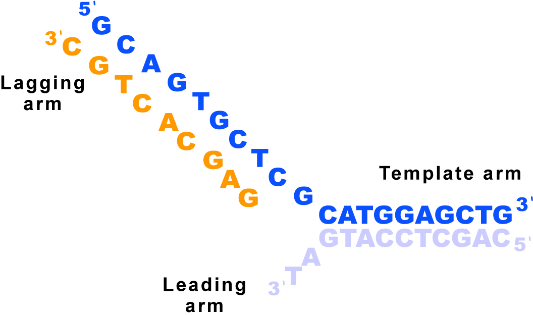
| 07:20, 29 November 2009 | Dna three way junction sequence.jpg (file) |  |
182 KB | DNA sequence in the structure | 2 |
| 03:50, 28 November 2009 | 1gm5 showing chains ADP.png (file) |  |
609 KB | Made with Jmol | 1 |
| 05:41, 2 November 2009 | 1s72ALPHACplusccapuro.PDB.gz (file) | |
154 KB | 1s72 where nucleic and proteins just phosphorus and alpha carbon backbone but where i added back CCA-puro from 1ffz as chain 5 | 1 |
| 02:27, 2 November 2009 | 1s72simplified.PDB.gz (file) | |
314 KB | Ran Eric Martz's alphac 2.0 on 1s72 to simplify | 1 |
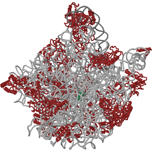
| 01:38, 2 November 2009 | Lsu side and front white background.png (file) |  |
292 KB | 1s72 from side and front using Swiss PDB viewer | 1 |
| 20:21, 1 November 2009 | 3ews3g0hJUSTSTARTEND.pdb (file) | |
126 KB | 3ews and 3g0h in a single file as they had been morphed by Yale Server. The intermediate models were removed and the extra atoms for 3ews added back in by Magicfit in Swiss PDB viewer. | 3 |
| 04:22, 1 November 2009 | 3g0hSIMPLER.jpg (file) |  |
257 KB | The default image Image:3g0h.png looked quite complex so I made a simpler one still in the style of many of the default images. | 1 |
| 01:21, 1 November 2009 | 3ewsBIOLUNIT1.pdb.gz (file) | |
141 KB | Biological unit 1 since asymmetric unit has 2 | 1 |
| 23:13, 31 October 2009 | 3ews3g0hmorphBETTER.pdb.gz (file) | |
484 KB | Morph of 3ews to 3g0h. Improved by adding back the ligands and RNA and the extra atoms to model 1 that came from using magicFit to translate it over automatically in Swiss-PDB Viewer | 1 |
| 04:49, 31 October 2009 | 3ews3g0hmorphFIXED.pdb.gz (file) | |
482 KB | See Image:3ews3g0hmorph.pdb.gz because I don't feel like entering all the information again since I had to spend time fixing this and don't know if it is done yet. | 1 |
| 04:31, 31 October 2009 | 3ews3g0hmorph.pdb.gz (file) | |
482 KB | morph of 3ews to 3g0h gzipped No copyright since PDB says pdb files and derived files aren't protected. | 1 |
| 03:01, 23 October 2009 | 3fpntorotatedversion.pdb (file) | |
1.01 MB | 3fpn normal view morphed to view like in Figure 3 of paper describing structure. There is no copyright as PDB says no copyright on PDB files or files derived from them. | 1 |
| 20:24, 18 October 2009 | 2eyqBIOLUNIT.pdb (file) | |
751 KB | Biological Unit of 2eyq. No copyright needed since the PDB says PDB files and those derived do not fall under copyrights. | 1 |
| 20:25, 8 October 2009 | SSU front and LSU front side by side white backgroundSMALL.jpg (file) |  |
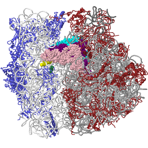
313 KB | SSU from Ramakrishnan work with A-site Anticodon Stem Loop bound as well as mRNA analaog show. LSU from Steitz and Moore labs work shown. Derived from PDB files available at Protein Data Bank which says they have no copyright if you use derived structur | 1 |
| 20:05, 8 October 2009 | Screen capture of 70S white background spacefill tRNAS.jpg (file) |  |
1 MB | screen capture (most anything else causes crash) of 70S ribosome from Pymol. This is the Noller 2.8 Angstrom structure (2j00 and 2j01) with the A-site tRNA from the 5.5 Angstrom (1gix) fit back in with the portion seen at 2.8 Angstrom. According to Prote | 1 |
| 00:59, 2 October 2009 | 3dmqBIOLUNIT.pdb (file) | |
640 KB | biological molecule 1 of 3dmq from https://www.pdb.org/pdb/explore/explore.do?structureId=3DMQ From the PDB "Data files contained in the PDB archive (ftp://ftp.wwpdb.org) are free of all copyright restrictions and made fully and freely available for both | 1 |
| 05:39, 27 September 2009 | 2db3BIOMOL.pdb (file) | |
322 KB | biological molecule 1 of 2db3 from https://www.rcsb.org/pdb/explore/explore.do?structureId=2DB3 From the PDB "Data files contained in the PDB archive (ftp://ftp.wwpdb.org) are free of all copyright restrictions and made fully and freely available for both | 1 |
| 04:47, 27 September 2009 | 1r9fTRUBIOUNIT.pdb (file) | |
238 KB | biological unit of 1r9f from https://www.rcsb.org/pdb/files/1R9F.pdb1.gz (REMOVED THE MODEL NUMBERS AND SECOND RNA and made second A chain D using a python script (SEE BELOW) From the PDB "Data files contained in the PDB archive (ftp://ftp.wwpdb.org) ar | 1 |
| 04:28, 27 September 2009 | 1r9fTRUEBIOLUNIT.pdb (file) | |
239 KB | biological unit of 1r9f from https://www.rcsb.org/pdb/files/1R9F.pdb1.gz From the PDB "Data files contained in the PDB archive (ftp://ftp.wwpdb.org) are free of all copyright restrictions and made fully and freely available for both non-commercial and co | 1 |
| 04:12, 27 September 2009 | 1r9f biolunit.pdb (file) | |
301 KB | biological unit of 1r9f from https://www.rcsb.org/pdb/files/1R9F.pdb1.gz | 1 |
| 03:05, 20 April 2009 | 1l8bmonomer.pdb (file) | |
158 KB | Just one monomer of the Messenger RNA 5' Cap-binding Protein (eIF4E) bound to 7-methylGpppG. The original file 1l8b had two in the unit cell of the crystal structure, in contrast to the biological unit. From the PDB "Data files contained in the PDB archi | 1 |
| 05:55, 1 February 2009 | 2az0WithPolyview3D.png (file) |  |
309 KB | 2az0 with Polyview-3D Settings used: Data source: PDB code=2az0 Initial rotation around X axis: -85.5 Initial rotation around Y axis: -77.2 Initial rotation around Z axis: 38.1 Rendering program: PyMol Type of image: Static slide Background color Red fac | 1 |
| 22:48, 29 January 2009 | 1jgo1giy.gz.pdb (file) | |
296 KB | pdb files 1jgo and 1giy as one gzipped file for use with jmol See Yusupov MM, Yusupova GZ, Baucom A, Lieberman K, Earnest TN, Cate JH, and Noller HF. 2001. Crystal structure of the ribosome at 5.5 Å resolution. Science 292:883-896. Epub 2001 Mar 29. an | 1 |
| 21:41, 27 November 2008 | 2h9cmorph2h9d.pdb (file) | |
828 KB | morph of 2h9c to 2h9d a and b chains. Pyruvate of 2h9d model added back by magic fitting 2h9d to last model of morph and extracting the 204 and 205 set of pyr atoms. | 1 |
| 07:04, 27 November 2008 | 2h9dmorph8.pdb (file) | |
850 KB | 8 model morph of chains A and B of 2h9d to chains C and D of 2h9d with pyruvates placed using magic fit of A and B part to first model and C and D part to last model. | 1 |
| 23:13, 25 November 2008 | 1atu1ezxwd.pdb (file) | |
8.06 MB | Daniel Seeman's file with just model numbers fixed by find and replace | 1 |
| 22:54, 25 November 2008 | 1atu 1ezxwd30.pdb (file) | |
4.03 MB | models 1-30 of Daniel Seeman's morph | 1 |
| 21:01, 25 November 2008 | Dsmorph.pdb (file) | |
1.61 MB | Models 1-6 of Daniel Seeman's morph | 1 |
| 20:57, 25 November 2008 | Ds models1-6.pdb (file) | |
1.61 MB | Models 1-6 of Daniel Seeman's morph | 1 |
| 20:23, 25 November 2008 | Me cropped smaller at Eileen Jewel performance 11-9-08..jpg (file) |  |
1,001 KB | Wayne Decatur in front row at Eilen Jewell private show November 9th 2008 | 1 |
| 04:26, 21 November 2008 | Green1ema.png (file) |  |
287 KB | GFP (1ema) made with POLYVIEW-3D Settings used: Data source: PDB code=1ema Initial rotation around X axis: -155.3 Initial rotation around Y axis: -54.6 Initial rotation around Z axis: 15.3 Rendering program: PyMol Type of image: Static slide Background co | 1 |
| 05:46, 9 November 2008 | Tetramer2zi0.pdb (file) | |
315 KB | 1 | |
| 05:41, 9 November 2008 | 2zi0 tetramer.pdb (file) | |
315 KB | 2 | |
| 02:35, 4 November 2008 | 2zioWithPolyview3D.png (file) |  |
174 KB | official 2zi0 fancy image made with [https://polyview.cchmc.org/polyview3d.html POLYVIEW-3D]<br> Settings used:<br> Data source: PDB code=2zi0 Initial rotation around X axis: 1.8 Initial rotation around Y axis: 40.0 Initial rotation around Z axis: 125.2 Re | 1 |
| 05:50, 21 October 2008 | Unremediated1rpuWithPolyview3D.png (file) |  |
345 KB | Unremediated 1rpu fancy image made with Polyview-3D Settings used:</h3> Data source: <b>Custom file</b><br> Structure source: <b>OTHER</b> server<br> Show ligands<br> Ligands are rendered as <b>spheres</b><br> Initial rotation around <b>X</b> axis: <b>115 | 1 |