File:Dimerization site GSS.jpg
Original file (1,050 × 689 pixels, file size: 390 KB, MIME type: image/jpeg)
Proteopedia Page Contributors and Editors (what is this?)
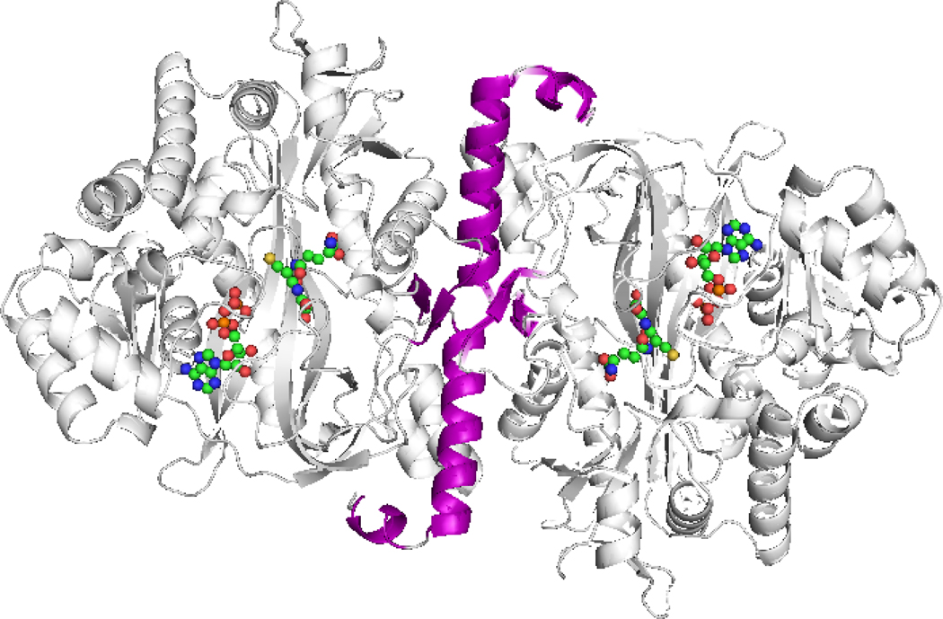
Shown in purple is the site of dimerization formed by the two homogenous protein subunits.
Image borrowed from the NCBI databank for non-profit and educational purposes only. I claim no credit for the image shown. All credit goes to Slavens, et al (2011) at the following article:
Slavens KD, Brown TR, Barakat KA, Cundari TR, Anderson ME. 2011. Valine 44 and valine 45 of human glutathione synthetase are key for subunit stability and negative cooperativity. Biochem & Biophys Resear Comm, 410(3): 597-601. doi: 10.1016/j.bbrc.2011.06.034
Proteopedia Page Contributors and Editors (what is this?)
File history
Click on a date/time to view the file as it appeared at that time.
| Date/Time | Thumbnail | Dimensions | User | Comment | |
|---|---|---|---|---|---|
| current | 01:09, 5 December 2013 | | 1,050 × 689 (390 KB) | Elliott Wyatt (talk | contribs) | Shown in purple is the site of dimerization formed by the two homogenous protein subunits. |
You cannot overwrite this file.
File usage
The following page uses this file:


