Uploads by Israel Hanukoglu
From Proteopedia
Jump to navigationJump to search
This special page shows all uploaded files.
| Date | Name | Thumbnail | Size | Description |
|---|---|---|---|---|
| 10:09, 16 August 2015 | NAD-NADH-redox-reactions.png (file) |  |
43 KB | This image shows the redox reactions of NAD together with the structures of the oxidized (NAD+) and reduced (NADH) forms. |
| 15:54, 23 July 2014 | 3-phosphoglycerate dehydrogenase-2P9E-sheet.png (file) |  |
399 KB | 3-phosphoglycerate dehydrogenase (2P9E) beta sheet in the NAD binding domain. The two beta-strands that form the core of the Rossmann fold are marked in dark-blue color. |
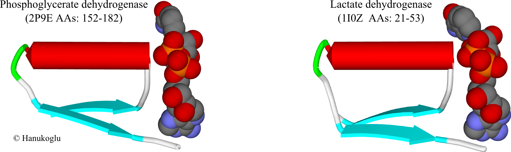
| 15:14, 23 July 2014 | NAD-binding-sites.png (file) |  |
283 KB | NAD binding sites of 3-phosphoglycerate dehydrogenase (2P9E) and lactate dehydrogenase (1I0Z). NAD is shown in CPK mode. |
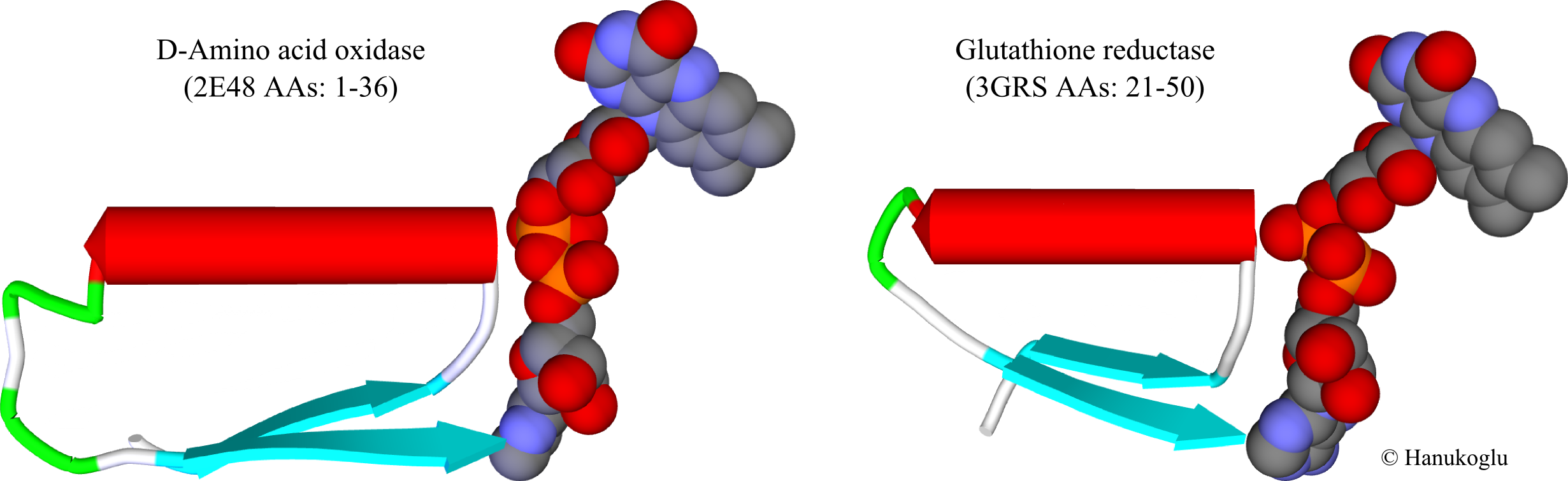
| 14:54, 23 July 2014 | FAD-binding-sites.png (file) |  |
394 KB | FAD binding sites of D-amino acid oxidase (2E48) and glutathione reductase (3GRS). FAD is shown in CPK mode. |
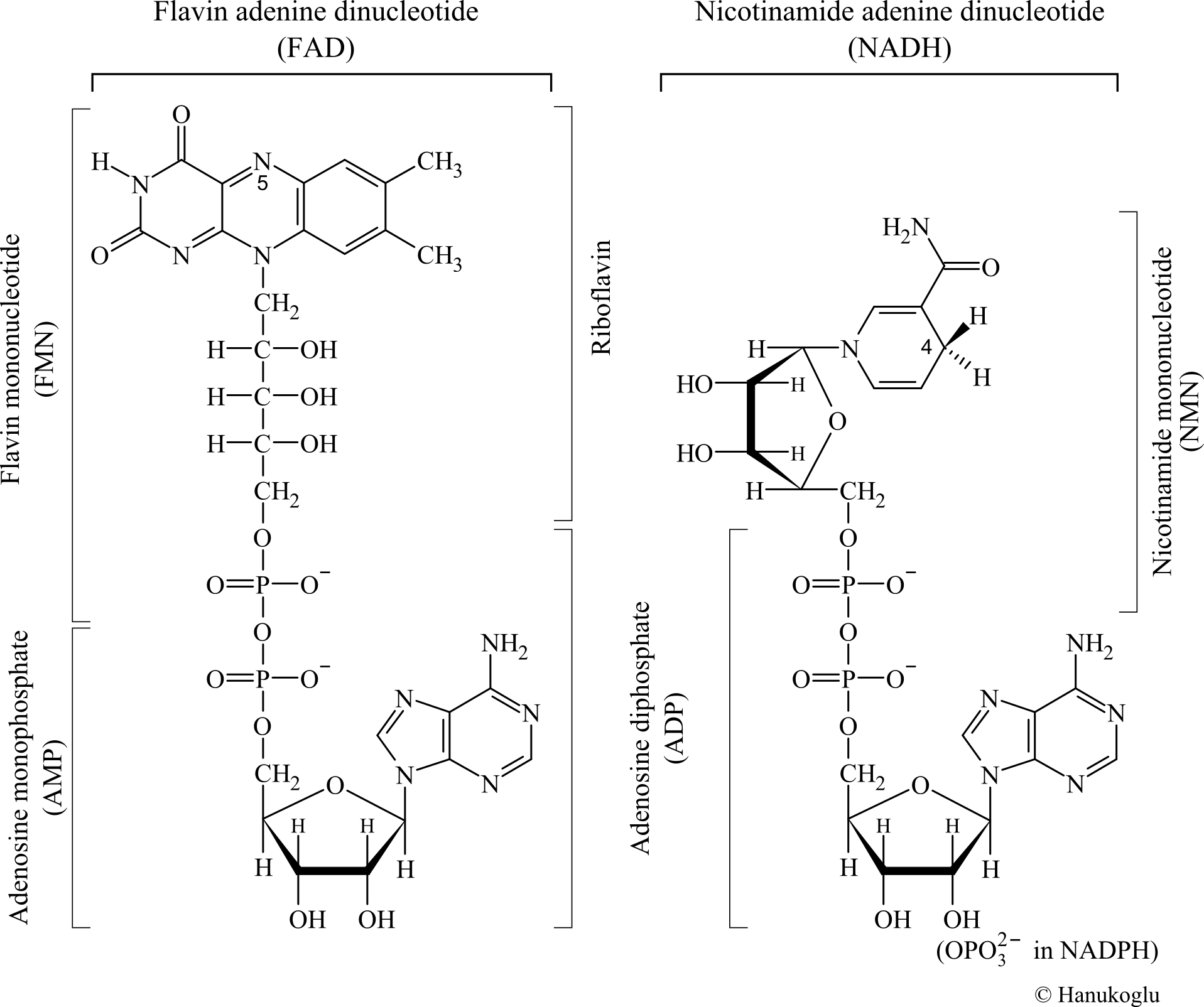
| 13:56, 23 July 2014 | FAD-NADH-Structures.png (file) |  |
210 KB | Structures of dinucleotides FAD and NADH in vertical orientation. Prepared by Israel Hanukoglu. |