File:Figure 1.JPG
From Proteopedia
No higher resolution available.
Figure_1.JPG (225 × 330 pixels, file size: 14 KB, MIME type: image/jpeg)
Proteopedia Page Contributors and Editors (what is this?)
FIGURE 1: Movement of Protons (H+) Across Inner Mitochondrial Membrane 1.The electron transport chain pumps protons (H+) into the inter membrane space. 2.Protons (H+) in the matrix bond with oxygen (O2) to form water (H2O) in a reduction reaction. 3.Protons (H+) from the inter membrane space are used by ATP synthase, to provide energy for the conversion of ADP to ATP.
Proteopedia Page Contributors and Editors (what is this?)
File history
Click on a date/time to view the file as it appeared at that time.
| Date/Time | Thumbnail | Dimensions | User | Comment | |
|---|---|---|---|---|---|
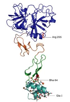
| current | 14:58, 22 April 2013 | | 225 × 330 (14 KB) | Student (talk | contribs) | Figure 1. 3-D structure of Protein Z: N-terminal Gla domain (light blue), EGF2 domain (green), EGF1 domain (orange), SP domain (dark blue). [5] |
| 21:28, 29 May 2012 |  | 491 × 351 (18 KB) | Deepti Ajjampore (talk | contribs) | FIGURE 1: Movement of Protons (H+) Across Inner Mitochondrial Membrane 1.The electron transport chain pumps protons (H+) into the inter membrane space. 2.Protons (H+) in the matrix bond with oxygen (O2) to form water (H2O) in a reduction reaction. 3.Proto |
You cannot overwrite this file.
File usage
The following page uses this file:


