File:Figure 4.JPG
From Proteopedia
No higher resolution available.
Figure_4.JPG (332 × 350 pixels, file size: 19 KB, MIME type: image/jpeg)
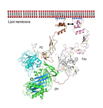
Figure 4. Model of the ternary complex of PZ-ZPI-FXa on the phospholipid membrane surface. PZ- N-terminal domains Gla, EGF1, and EGF2 (brown). ZPI (green) with reactive loop (red) and β-sheet A (light blue). FXa- N-terminal domains Gla, EGF1, and EGF2 (pink). Ca2+ (blue dots). Hydrophilic phosphate groups of phospholipids (red dots), hydrophobic tails (black dashes). Disulfide bonds (yellow sticks). Interaction of PZ and FXa Gla domains (black arrows). [4]
File history
Click on a date/time to view the file as it appeared at that time.
| Date/Time | Thumbnail | Dimensions | User | Comment | |
|---|---|---|---|---|---|
| current | 15:13, 22 April 2013 | | 332 × 350 (19 KB) | Student (talk | contribs) | Figure 4. Model of the ternary complex of PZ-ZPI-FXa on the phospholipid membrane surface. PZ- N-terminal domains Gla, EGF1, and EGF2 (brown). ZPI (green) with reactive loop (red) and β-sheet A (light blue). FXa- N-terminal domains Gla, EGF1, and EGF2 (p |
You cannot overwrite this file.
File usage
The following page uses this file:


