File:Snare complex.jpg
Original file (588 × 737 pixels, file size: 71 KB, MIME type: image/jpeg)
Proteopedia Page Contributors and Editors (what is this?)
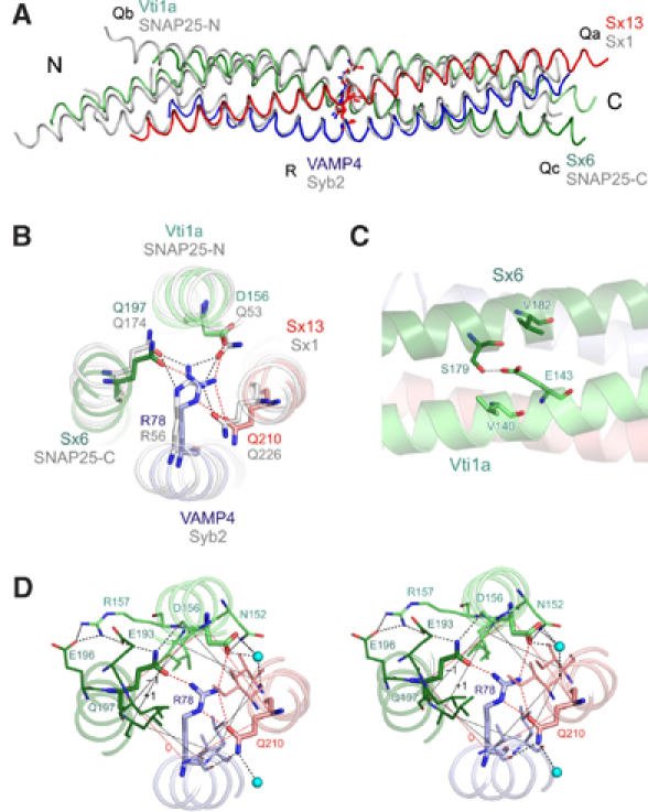
Crystal structure of the early endosomal SNARE complex. (A) Backbone overlay of the structures of the early endosomal SNARE complex (in color) and the structure of the neuronal SNARE complex (gray). QabcR classification is indicated next to protein names. (B) N- to C-terminal view of the 0 layer containing the unusual aspartate in vti1a (ribbon-ball-and-stick representation). Color, early endosomal SNARE complex; gray, neuronal SNARE complex. (C) Residue E143 builds a salt bridge with the residue S179. The residue E143 is also sandwiched by residues V182 and V140, which strengthen polar interactions. In the late endosomal and in the neuronal SNARE complex, this glutamate is conserved and interacts with a conserved arginine that occupies the position equivalent to V182 (Antonin et al, 2002; Ernst and Brunger, 2003a). (D) Stereo view of the 0 layer, including hydrogen bonds with surface residues and water molecules.
Proteopedia Page Contributors and Editors (what is this?)
File history
Click on a date/time to view the file as it appeared at that time.
| Date/Time | Thumbnail | Dimensions | User | Comment | |
|---|---|---|---|---|---|
| current | 13:26, 22 December 2013 | | 588 × 737 (71 KB) | Camille Roesch (talk | contribs) | Crystal structure of the early endosomal SNARE complex. (A) Backbone overlay of the structures of the early endosomal SNARE complex (in color) and the structure of the neuronal SNARE complex (gray). QabcR classification is indicated next to protein names. |
You cannot overwrite this file.
File usage
The following 2 pages use this file:


