Click here to color the tRNA 3D structure to match the secondary structure above.
The acceptor stem includes the 5' and 3' ends of the tRNA. The 5' end is generated by RNaseP :-). The 3' end is the site which is charged with amino acids for translation. Some aminoacyl tRNA synthetases interact with both the acceptor 3' end and
the anticodon when charging tRNAs. Note how far the 3' end is from the anticodon loop, at bottom, by clicking here. Note also how the acceptor stem stacks onto the TpsiC stem to form a continuous helix. The anticodon stem also stacks onto the junction between the variable loop and the D stem to form another nearly perfect helix. The TpsiC and D loops interact to bring the "cloverleaf" secondary structure in to the L-shaped tertiary structure.
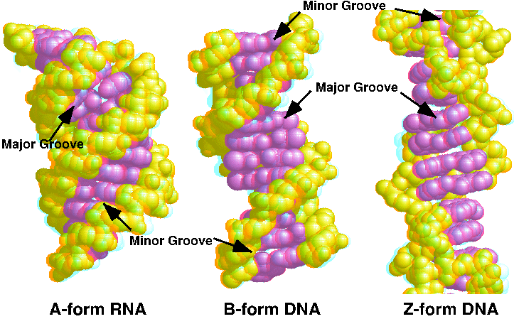
The tour starts with the Default view. Now look at this space filling view.The backbone is yellow and the bases are magenta. Note that the major groove (at the top, when you have just clicked the button) is very deep.
Now change the display to make the show the sugar-phosphate backbone as pseudo-bonds connecting the phosphate atoms. Now the bases are easier to see. Notice how they are stacked upon each other but not perpendicular to the axis of the double helix. They are also displaced to the side of the axis. The result is a wide, short helix. Note also that the backbone forms a smooth, continuous curve.
Each base pair stacks on the next similarly, as shown from this top view. This is the same top view of just the bases.
B-DNA stacks similarly, but compare this with Z-DNA, which behaves much differently. Essentially all helical RNA is in A form, but DNA can also be found in A form under certain conditions (particularly in RNA-DNA hybrids). The 2'-OH of ribose favors the C3'-endo sugar pucker necessary for A-form geometry. The O2' is easily seen as white spheres in this space fill view.
Forms of DNA shows a side-by-side comparison of A, B, and Z forms of DNA.
An interactive tutorial on DNA Structure, disponible también en español and eight other languages.
References
JSmol in Proteopedia [1] or to the article describing Jmol [2] to the rescue.
↑Hanson, R. M., Prilusky, J., Renjian, Z., Nakane, T. and Sussman, J. L. (2013), JSmol and the Next-Generation Web-Based Representation of 3D Molecular Structure as Applied to Proteopedia. Isr. J. Chem., 53:207-216. doi:https://dx.doi.org/10.1002/ijch.201300024
↑Herraez A. Biomolecules in the computer: Jmol to the rescue. Biochem Mol Biol Educ. 2006 Jul;34(4):255-61. doi: 10.1002/bmb.2006.494034042644. PMID:21638687 doi:10.1002/bmb.2006.494034042644
Proteopedia Page Contributors and Editors (what is this?)