Welcome to Proteopedia ISSN 2310-6301The free, collaborative 3D-encyclopedia of proteins & other molecules
Selected Pages
Art on Science
Journals
Education
Coronavirus Spike Protein Priming
by Eric Martz
Coronavirus SARS-CoV-2 (responsible for COVID-19) has a spike protein on its surface, which enables it to infect host cells. Initially, proteases in the lungs clip the homo-trimeric spike protein at a unique sequence. This primes it, causing it to extend its receptor binding surface (shown in the above animation), optimizing binding to the host cell's ACE2 receptor (not shown). Next, spike protein initiates fusion of the virus and host cell membranes (not shown), enabling the virus RNA to enter the cell and initiate production of new virions. Knowledge of spike protein's molecular structure and function is crucial to developing effective therapies and vaccines.
>>> Visit this page >>>
Opening a Gate to Human Health
by Alice Clark (PDBe)
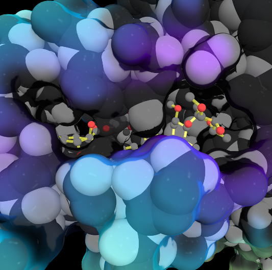
In the 1970s, an exciting discovery of a family of medicines was made by the Japanese scientist Satoshi Ōmura. One of these molecules, ivermectin, is shown in this artwork bound in the ligand binding pocket of the Farnesoid X receptor, a protein which helps regulate cholesterol in humans. This structure showed that ivermectin induced transcriptional activity of FXR and could be used to regulate metabolism.
Structure of Anticancer Ruthenium Half-Sandwich Complex Bound to Glycogen Synthase Kinase 3ß
G Atilla-Gocumen, L Di Costanzo, E Meggers. J Biol Inorg Chem. 2010 doi: 10.1007/s00775-010-0699-x
A crystal structure of an organometallic half-sandwich ruthenium complex bound to glycogen synthase kinase 3ß (GSK-3ß) reveals that the inhibitor binds to the ATP binding site via an induced fit mechanism utilizing several hydrogen bonds and hydrophobic interactions. Importantly, the metal is not involved in any direct interaction with the protein kinase but fulfills a purely structural role.
The Capsid of a virus is its outer shell or "skin". Viruses have evolved intricate and elegant ways to assemble
capsid protein chains into complete, usually spherical capsids, often with icosahedral symmetry. Pictured is an extremely simplified model of a capsid, where a single enlarged atom represents each of the 360 protein chains in the capsid of the Simian Virus 40 (SV40), a member of a group of cancer-causing viruses that has been extensively researched for decades.