Main Page

From Proteopedia
Revision as of 12:08, 21 October 2018 by Joel L. Sussman (talk | contribs)
Jump to navigationJump to search
ISSN 2310-6301

As life is more than 2D, Proteopedia helps to bridge the gap between 3D structure & function of biomacromolecules

Often it is difficult to utilize the wealth of information found in 3D biomacromolecular structures. Proteopedia's goal is to present structure/function information on these molecules in a user-friendly manner to a broad scientific audience.


Selected Pages Art on Science Journals Education
HIV-1 protease

by David Canner
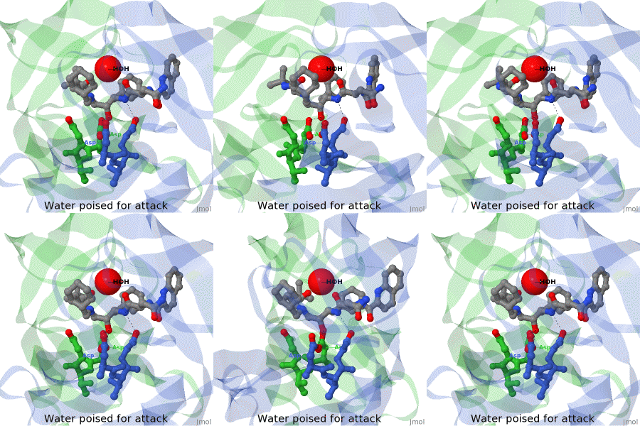
The X-ray structure of HIV-1 protease reveals that it is composed of two symmetrically related subunits which form a tunnel where they meet. This is critical because it contains the active site of the protease, consisting on two Asp-Thr-Gly conserved sequences, making it a member of the aspartyl protease family. The two catalytic Asp's either interact with the incoming water or protonate the carbonyl to make the carbon more electrophilic for the incoming water.

>>> Visit this page >>>

Molecular Sculpture

by Eric Martz
A historical review on sculptures and physical models of macromolecules.

>>> Visit this page >>>

Structure of Anticancer Ruthenium Half-Sandwich Complex Bound to Glycogen Synthase Kinase 3ß

G Atilla-Gocumen, L Di Costanzo, E Meggers. J Biol Inorg Chem. 2010 doi: 10.1007/s00775-010-0699-x
A crystal structure of an organometallic half-sandwich ruthenium complex bound to glycogen synthase kinase 3ß (GSK-3ß) reveals that the inhibitor binds to the ATP binding site via an induced fit mechanism utilizing several hydrogen bonds and hydrophobic interactions. Importantly, the metal is not involved in any direct interaction with the protein kinase but fulfills a purely structural role.

>>> Visit this I3DC complement >>>

Touch-Sensitive Channel

Touching stretches cell membranes, opening mechanosensitive ion channels, leading to sensation by the nervous system. Pictured is the transmembrane region of a similar channel in bacteria. When closed, the narrow opening is lined by hydrophobic amino acid sidechains, making it non-conductive to ions.

>>> See more animations and explanation >>>

How to add content to Proteopedia

Video Guides

Who knows ...

List of Art on Science pages

About Interactive 3D Complements - I3DCs

List of I3DCs

How to get an I3DC for your paper

Teaching strategies using Proteopedia

Examples of pages for teaching

How to add content to Proteopedia

About Contact Table of Contents Structure Index Help

Proteopedia Page Contributors and Editors (what is this?)

Jaime Prilusky, Joel L. Sussman, Angel Herraez