Image:Domain phenylketonuria 4.jpg
From Proteopedia

Size of this preview: 600 × 600 pixels
Full resolution (903 × 903 pixel, file size: 282 KB, MIME type: image/jpeg)
Summary
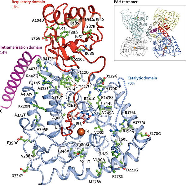
This is a 3D structure of a PheOH subunit showing its three domains, regulatory, catalytic, and tetramerization. Also many of the possible mutations are labeled along the chain giving a perspective of where the most mutations occur. There is also a picture of the tetrameric structure.
Licensing
{{subst:No license from license selector|Somewebsite}}
File history
Click on a date/time to view the file as it appeared at that time.
| Date/Time | User | Dimensions | File size | Comment | |
|---|---|---|---|---|---|
| (current) | 00:43, 3 May 2012 | Stefan Phelan (Talk | contribs) | 903×903 | 282 KB | This is a 3D structure of a PheOH subunit showing its three domains, regulatory, catalytic, and tetramerization. Also many of the possible mutations are labeled along the chain giving a perspective of where the most mutations occur. There is also a pictur |
- Edit this file using an external application
See the setup instructions for more information.
Links
The following pages link to this file:
