Image:PCSK9 domains2.png
From Proteopedia
Size of this preview: 800 × 450 pixels
Full resolution (1280 × 720 pixel, file size: 84 KB, MIME type: image/png)
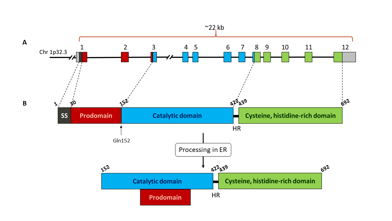
Schematic representation of PCSK9 gene (A) and protein (B). A: exons of PCSK9 gene are shown as coloured boxes. Each colour corresponds to a different domain of the protein. B: Representation of PCSK9 protein domains: signal sequence (SS, black), prodomain (red), catalytic domain (blue) and Cys, His rich C-terminal domain (green). The autocatalytic cleavage site at Gln152 is indicated with an arrow. Once the protein has been processed in the endoplasmic reticulum (ER), the prodomain remains bound to the catalytic domain. The catalytic domain is linked to the C-terminal domain through a 18 amino acids hinge region (HR). Numbers above each domain indicate the amino acid number of the protein sequence.
File history
Click on a date/time to view the file as it appeared at that time.
| Date/Time | User | Dimensions | File size | Comment | |
|---|---|---|---|---|---|
| (current) | 17:22, 30 December 2017 | Rafael Romero Becerra (Talk | contribs) | 1280×720 | 84 KB | Schematic representation of PCSK9 gene (A) and protein (B). A: exons of PCSK9 gene are shown as coloured boxes. Each colour corresponds to a different domain of the protein. B: Representation of PCSK9 protein domains: signal sequence (SS, black), prodomai |
- Edit this file using an external application
See the setup instructions for more information.
Links
The following pages link to this file:
