A-RNA tour
From Proteopedia
A-form RNA
Source [1] Structural highlights
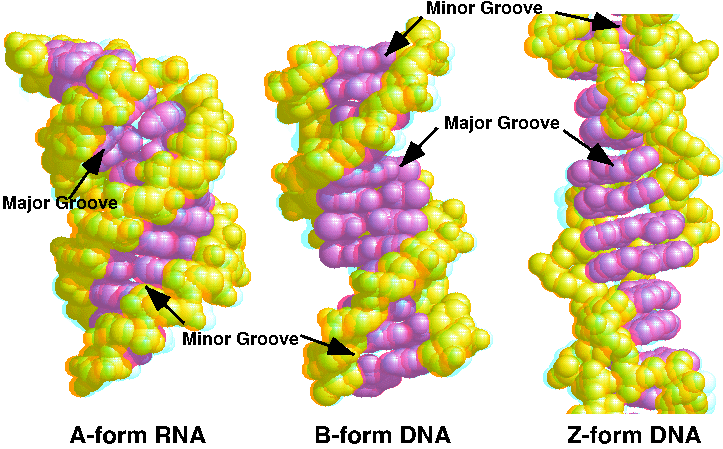
Take the TourThe tour starts with the Default view. Now look at this space filling view.The backbone is yellow and the bases are magenta. Note that the major groove (at the top, when you have just clicked the button) is very deep. Now change the display to make the show the sugar-phosphate backbone as pseudo-bonds connecting the phosphate atoms. Now the bases are easier to see. Notice how they are stacked upon each other but not perpendicular to the axis of the double helix. They are also displaced to the side of the axis. The result is a wide, short helix. Note also that the backbone forms a smooth, continuous curve. You can look at just four of the base pairs..You are looking into the major groove and the colors of the base pairs alternate. You can also look at just the bases. Each base pair stacks on the next similarly, as shown from this top view. This is the same top view of just the bases. B-DNA stacks similarly, but compare this with Z-DNA, which behaves much differently. Essentially all helical RNA is in A form, but DNA can also be found in A form under certain conditions (particularly in RNA-DNA hybrids). The 2'-OH of ribose favors the C3'-endo sugar pucker necessary for A-form geometry. The O2' is easily seen as white spheres in this space fill view. You can compare it with the DNA forms by looking at this 3D red-blue stereo picture of A, B, and Z DNA |
| ||||||||||

See Also
- Z-DNA model tour and Z-DNA
- B-DNA tour
- Transfer RNA tour
- A more general overview will be found at DNA.
- Forms of DNA shows a side-by-side comparison of A, B, and Z forms of DNA.
- An interactive tutorial on DNA Structure, disponible también en español and eight other languages.
References
JSmol in Proteopedia [2] or to the article describing Jmol [3] to the rescue.
- ↑ Dickerson RE, Drew HR, Conner BN, Wing RM, Fratini AV, Kopka ML. The anatomy of A-, B-, and Z-DNA. Science. 1982 Apr 30;216(4545):475-85. PMID:7071593
- ↑ Hanson, R. M., Prilusky, J., Renjian, Z., Nakane, T. and Sussman, J. L. (2013), JSmol and the Next-Generation Web-Based Representation of 3D Molecular Structure as Applied to Proteopedia. Isr. J. Chem., 53:207-216. doi:http://dx.doi.org/10.1002/ijch.201300024
- ↑ Herraez A. Biomolecules in the computer: Jmol to the rescue. Biochem Mol Biol Educ. 2006 Jul;34(4):255-61. doi: 10.1002/bmb.2006.494034042644. PMID:21638687 doi:10.1002/bmb.2006.494034042644
