Cystine
From Proteopedia
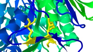
Cystine is formed by the oxidation of two proximal cysteine residues, covalently linking them in a disulfide bond.

Two cystines in human acid-beta-glucosidase (GlcCerase), the enzyme mutated in Gaucher disease, from 1ogs.
|
Contents |
Visualization
FirstGlance in Jmol displays all cystines (disulfide bonds) in any given protein in a clear manner. To display a protein in FirstGlance and see its disulfide bonds:
- Find a page in Proteopedia about that protein. You can use the search slot at left to search for the name of the protein, or a specific PDB code.
- If the page is not titled with a PDB code, look for mention of a PDB code, and go to the page titled with that PDB code.
- Click on the link FirstGlance beneath the molecule.
- Once you see the protein in FirstGlance, click on More Views.., then on Disulfide bonds: show all.
Eventually, you will be able to select and display disulfide bonds in Proteopedia's Scene Authoring Tools (SAT). In March, 2012, this is on the Wishlist. Currently, in the SAT, you can select CYS, which includes both cysteine and cystine.
Articles
Articles in Proteopedia concerning cystine include:
- Disulfide Connectivity of Velaglucerase, a.k.a. acid-beta-glucosidase (GlcCerase)
- Serine Proteases
- PHB synthase in Rhodobacter sphaeroides
- Altering Disulfide Bonds in a Structure Using PyMol
Cystine Categories
To view automatically seeded "Category" indices concerning cystine, see:
- Cyclic cystine knot
- Cystine knot superfamily
- Cystine knot
- Cystine stabilized alpha-beta motif
- Inhibitor cystine knot
- Cystine-stabilized alpha-helical motif
- Cystine-knot
- Cystine-rich
- Cystine-knot growth factor
- Inhibitor cystine knot motif
- Inhibitor cystine-knot
- Cyclic cystine knot motif
- Homodimer,cystine knot
- Inhibitory cystine knot
- Fad-cystine-oxidoreductase
- Cystine knot motif
References and Notes
See Also
Additional Literature
- Ladenstein R, Ren B. Reconsideration of an early dogma, saying "there is no evidence for disulfide bonds in proteins from archaea". Extremophiles. 2008 Jan;12(1):29-38. Epub 2007 May 17. PMID:17508126 doi:10.1007/s00792-007-0076-z
- Dvir H, Harel M, McCarthy AA, Toker L, Silman I, Futerman AH, Sussman JL. X-ray structure of human acid-beta-glucosidase, the defective enzyme in Gaucher disease. EMBO Rep. 2003 Jul;4(7):704-9. PMID:12792654 doi:10.1038/sj.embor.embor873
