Image:Fig 1.jpg
From Proteopedia
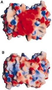
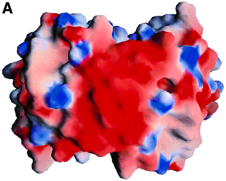
Fig. 1 shows the electrostatic surface potential map of A(HB19) and B(212). HB19 is a virulent strain from oMG A, and 212 is from a non-virulent strain. (Source: Kumaran et al. 2001)
File history
Click on a date/time to view the file as it appeared at that time.
| Date/Time | User | Dimensions | File size | Comment | |
|---|---|---|---|---|---|
| (current) | 17:51, 24 April 2012 | Gayatri Setia (Talk | contribs) | 177×303 | 11 KB | Fig. 1 shows the electrostatic surface potential map of A(HB19) and B(212). HB19 is a virulent strain from oMG A, and 212 is from a non-virulent strain. (Source: Kumaran et al. 2001) |
| 17:49, 24 April 2012 | Gayatri Setia (Talk | contribs) | 741×593 | 84 KB | Fig. 1 shows the electrostatic surface potential map of A(HB19) and B(212). HB19 is a virulent strain from oMG A, and 212 is from a non-virulent strain. (Source: Kumaran et al. 2001) |
- Edit this file using an external application
See the setup instructions for more information.
Links
The following pages link to this file:
