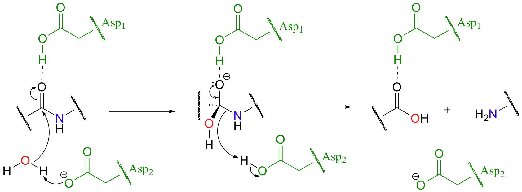
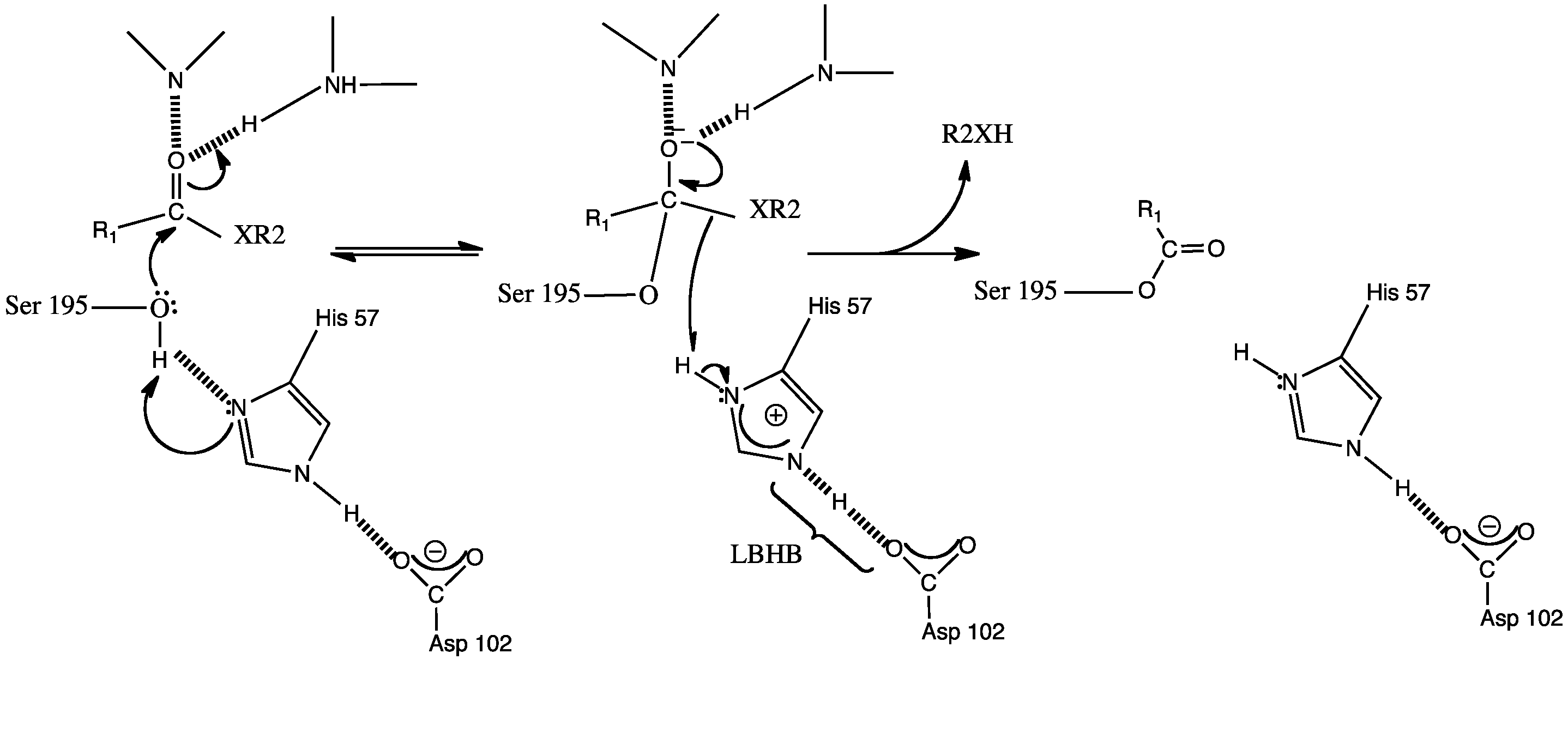
Image:Mechanism.png
From Proteopedia
| Error creating thumbnail: libgomp: Thread creation failed: Resource temporarily unavailable |
Size of this preview: 800 × 564 pixels
Full resolution (2182 × 1538 pixel, file size: 1.14 MB, MIME type: image/png)
Proteopedia Page Contributors and Editors (what is this?)
Charles Short, Joseph Gareis, Rushda Hussein, Hanan Busaileh, Zhichang Yang, Francis Ayombil, Jamie C. Gladfelder, Cory Tiedeman
File history
Click on a date/time to view the file as it appeared at that time.
| Date/Time | User | Dimensions | File size | Comment | |
|---|---|---|---|---|---|
| (current) | 18:57, 6 April 2023 | Rushda Hussein (Talk | contribs) | 2182×1538 | 1.14 MB | |
| 18:55, 6 April 2023 | Rushda Hussein (Talk | contribs) | 2182×1538 | 1.14 MB | ||
| 03:29, 18 April 2022 | Joseph Gareis (Talk | contribs) | 1478×762 | 294 KB | ||
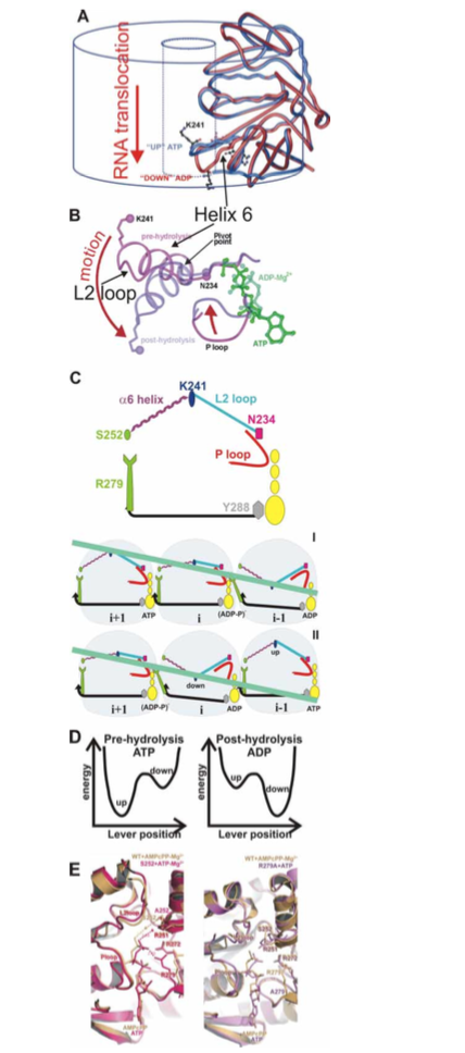
| 13:37, 17 December 2015 | Student (Talk | contribs) | 428×968 | 319 KB | (A–B) Loop L2 movement in the central channel (C) Mechanism of sequential hydrolysis of ATP (yellow) and RNA (light green) translocation for three subunits viewed from within the central | |
| 00:11, 27 November 2012 | Charles Short (Talk | contribs) | 1684×620 | 162 KB | mechanism | |
| 21:07, 25 November 2012 | Hanan Busaileh (Talk | contribs) | 647×260 | 51 KB | ||
| 21:07, 25 November 2012 | Hanan Busaileh (Talk | contribs) | 647×260 | 51 KB | ||
| 19:22, 19 November 2012 | Zhichang Yang (Talk | contribs) | 688×365 | 48 KB | ||
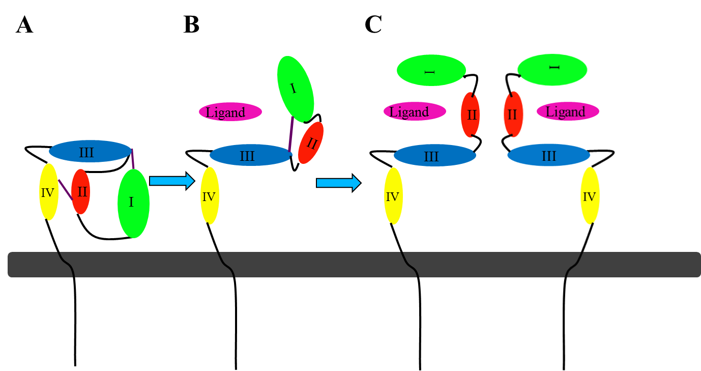
| 23:46, 6 November 2012 | Jamie C. Gladfelder (Talk | contribs) | 1378×729 | 55 KB | This picture illustrates the mechanism in which EGFR, HER3, and HER4 change conformation in order to dimerize and activate further cell signaling. A) Sub-domain I (green) forms an interaction (purple line) with sub-domain III (blue). Sub-domain II (red) f | |
| 23:21, 14 April 2012 | Francis Ayombil (Talk | contribs) | 2876×1354 | 124 KB | mech 1 | |
| 05:43, 1 March 2010 | Cory Tiedeman (Talk | contribs) | 978×379 | 59 KB |
- Edit this file using an external application
See the setup instructions for more information.
Links
The following pages link to this file:
