Image:Trichosurin ligand binding.jpg
From Proteopedia
No higher resolution available.
Trichosurin_ligand_binding.jpg (523 × 442 pixel, file size: 109 KB, MIME type: image/jpeg)
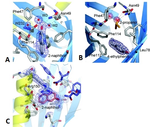
Trichosurin structure, topology and ligand binding
(A) A 2(Fo)−(Fc) electron density map contoured at 1σ depicted in blue mesh showing the 2-naphthol ligand bound within the trichosurin barrel and the hydrogen bonds made to water W3 and Asn49 ND1.
(B) 2(Fo)−(Fc) electron density map contoured at 1σ showing 4-ethylphenol (surrounded by electron density mesh) donating a hydrogen bond to Leu78 O and occupying the lower part of the trichosurin-binding pocket. A 2-propanol molecule (orange and red) is still bound in the upper part of the binding pocket along with the water molecules W2 and W3 (red spheres).
(C) The second binding site for 2-naphthol between the helix, the outside of the barrel and the dimer interface. The blue mesh is from a 2(Fo)−(Fc) electron density map contoured at 1σ. The thin blue lines and the purple residues are from the second molecule of the dimer. ncbi_file
File history
Click on a date/time to view the file as it appeared at that time.
| Date/Time | User | Dimensions | File size | Comment | |
|---|---|---|---|---|---|
| (current) | 21:32, 25 January 2017 | Mina Schneider (Talk | contribs) | 523×442 | 109 KB | Trichosurin structure, topology and ligand binding (A) A 2(Fo)−(Fc) electron density map contoured at 1σ depicted in blue mesh showing the 2-naphthol ligand bound within the trichosurin barrel and the hydrogen bonds made to water W3 and Asn49 ND1. ( |
- Edit this file using an external application
See the setup instructions for more information.
Links
The following pages link to this file:
Metadata
This file contains additional information, probably added from the digital camera or scanner used to create or digitize it. If the file has been modified from its original state, some details may not fully reflect the modified image.
| Orientation | Normal |
|---|
