Nitrile hydratase
From Proteopedia
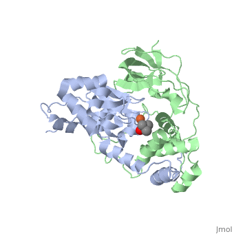
FunctionNitrile hydratase (NH) catalyze the hydration of nitriles to amides. NH contains Fe or Co ion in its active site. NH consists of two subunits: α and β and exists as dimer or tetramer with one metal ion bound per dimer. RelevanceNitrile hydratase is used industrially in the production of acrylamide and nicotinamide. Structural highlightsCrystal structure of the CN-hydrolase SA0302 from the pathogenic bacterium Staphylococcus aureus belonging to the Nit and NitFhit Branch of the nitrilase superfamily [1]The nitrilase, superfamily of protein enzyme, has in total over 180 known members. It includes a variety of thiol amidase enzymes involved in biosynthesis in plants, animals, fungi and prokaryotes [2]. All members of this superfamily have conserved the active site residues Glu-Lys-Cys, believed to form a catalytic triad. The consensus sequences flanking the catalytic residues supply the conserved motifs distinctive for all of the branches [2]. The superfamily can be classified into 13 branches, nine of which have known or predicted specificity for nitrilase, amidase, and CN-hydrolase reactions. Although the entire family has been considered “nitrilase-related”, only members of Branch 1 have demonstrated nitrilase activity. The remaining branches include enzymes with amidase or amide-condensation activity including aliphatic amidase, amino-terminal amidase, biotinidase, β-ureidopropionase, carbamylase, prokaryotic and eukaryotic NAD-synthetase, and apo-lipoprotein N-acyltransferase [3]. Analysis of the sequences and structures of CN-hydrolases with known three-dimensional structures shows that SA0302 definitely is a member of Branch 10 (Nit and NitFhit) of the nitrilase superfamily. Enzyme activities and substrate specificities of members of this branch are not yet characterized, in contrast to those of the members of Branches 1 to 9. The monomer consists of N-subdomain is colored in salmon and C-subdomain is in magenta. The active site is formed by the residues from both the N- and C-terminal sub-domains (catalytic residues are labeled). The protein contains 261 amino acids and consists of a four-layer αββα sandwich (color-coding: Alpha Helices, Beta Strands , Turns), consistent with the expected CN-hydrolase fold. The dimer, which was found in crystal and solution media, contains two accessible to the solvent active sites. First monomer is colored in green, second monomer is in darkmagenta, positions of catalytic triad residues are in red, and the conserved α6-β10 hairpin is shown in orange. The sequence alignments of SA0302 with four known nitrilase structures are presented in the image below.  They clearly show the conserved catalytic triad Glu41-Lys110-Cys146 which believed to describe the enzyme active site. These catalytic residues are located within three semi-conserved regions corresponding to their relative locations in the SA0302 protein sequence: region 1 – flanking E41; region 2 – following K110; and region 3 – flanking C146, which also has a protein signature consistent with the 21-residue “nucleophile elbow” motif [4]. These flanking catalytic sequences of SA0302 are in good accordance with consensus sequences of CN-hydrolase of Branch 10 (Nit and NitFhit) of the nitrilase superfamily [2]. On the basis of semi-conserved regions flanking the catalytic triad, we have assigned ten other uncharacterized protein sequences to Branch 10 of the nitrilase superfamily (see image below).  General view of the active site with the catalytic triad. Detailed analysis shows that active site includes five participants: catalytic triad Glu41-Lys110-Cys146, water molecule W272, and additional residue Glu119. In spite of growing interest about the details of the enzymatic mechanism of the members of Branch 10, at present little is known about the specificity of possible substrates or inhibitors. This is a very challenging biochemical problem that is still far from being resolved. At the moment, we can cite one important related reference [5]. In this paper, two murine nitrilases, including Nit1 and Nit2, were identified as targets for a dipeptide-chloroacetamide activity-based probe. The gel analysis of binding of Nit with these probes shows definite selectivity of labeling inside the Nit subfamily. Experimental data of positive labeling are as follows: Nit1: Leu-Tyr, Leu-His, Leu-Leu, Asp-Leu, Glu-Leu, Tyr-Leu, D-Leu-Asp, Leu-D-Asp, D-Leu-D-Asp, D-Leu-D-Asp Nit2: Leu-Tyr, Leu-His, Leu-Leu, Leu-Arg, Leu-Glu, Leu-Asp, D-Leu-Asp, where common dipeptide α-CA probes are in bold. These data implicate the specificity of substrate binding for a definite member of Branch 10 of the Nit-nitrilase superfamily. The present solution of the substrate-free bacterial protein structure of SA0302 gives the basis for substrate binding studies with the potential substrates or inhibitors. Analyzing the Catalytic Role of Active Site Residues in the Fe-Type Nitrile Hydratase from Comamonas testosteroni Ni1 [6]Active site of wild-type Fe-type nitrile hydratase from Comamonas testosteroni Ni1 (CtNHase, PDB ID: 4fm4) in ball-and-stick form is shown. A strictly conserved active site arginine residue (αR157) and two histidine residues (αH80 and αH81) located near the active site of the CtNHase, were mutated. The wild-type is in dark magenta, αH80A/αH81A mutant is in green, αH80W/αH81W is in orange, and the αR157A mutant is in deep sky blue. Click here to see animation of this scene. These mutant enzymes were examined for their ability to bind iron and hydrate acrylonitrile. For the αR157A mutant, the residual activity (kcat = 10 ± 2 s−1) accounts for less than 1 % of the wild-type activity (kcat = 1100 ± 30 s-1) while the Km value is nearly unchanged at 205 ± 10 mM. On the other hand, mutation of the active site pocket αH80 and αH81 residues to alanine resulted in enzymes with kcat values of 220 ± 40 and 77 ± 13 s-1, respectively, and Km values of 187 ± 11 and 179 ± 18 mM. The double mutant (αH80A/αH81A) was also prepared and provided an enzyme with a kcat value of 132 ± 3 s-1 and a Km value of 213 ± 61 mM. These data indicate that all three residues are catalytically important, but not essential. X-ray crystal structures of the αH80A/αH81A, αH80W/αH81W, and αR157A mutant CtNHase enzymes were solved to 2.0, 2.8, and 2.5 Å resolutions, respectively. In each mutant enzyme, hydrogen-bonding interactions crucial for the catalytic function of the αCys104-SOH ligand are disrupted. Disruption of these hydrogen bonding interactions likely alters the nucleophilicity of the sulfenic acid oxygen and the Lewis acidity of the active site Fe(III) ion. Superposition of the αβ heterodimer, the regions with pronounced difference between the wild-type and the mutant enzymes are indicated. Click here to see animation of this scene. |
| |||||||||||
3D structures of nitrile hydratase
Updated on 18-July-2023


References
- ↑ Gordon RD, Qiu W, Romanov V, Lam K, Soloveychik M, Benetteraj D, Battaile KP, Chirgadze YN, Pai EF, Chirgadze NY. Crystal structure of the CN-hydrolase SA0302 from the pathogenic bacterium Staphylococcus aureus belonging to the Nit and NitFhit Branch of the nitrilase superfamily. J Biomol Struct Dyn. 2013 Oct;31(10):1057-65. doi: 10.1080/07391102.2012.719111. , Epub 2012 Oct 2. PMID:23607706 doi:http://dx.doi.org/10.1080/07391102.2012.719111
- ↑ 2.0 2.1 2.2 Pace HC, Brenner C. The nitrilase superfamily: classification, structure and function. Genome Biol. 2001;2(1):REVIEWS0001. Epub 2001 Jan 15. PMID:11380987
- ↑ Brenner C. Catalysis in the nitrilase superfamily. Curr Opin Struct Biol. 2002 Dec;12(6):775-82. PMID:12504683
- ↑ Kumaran D, Eswaramoorthy S, Gerchman SE, Kycia H, Studier FW, Swaminathan S. Crystal structure of a putative CN hydrolase from yeast. Proteins. 2003 Aug 1;52(2):283-91. PMID:12833551 doi:http://dx.doi.org/10.1002/prot.10417
- ↑ Barglow KT, Saikatendu KS, Bracey MH, Huey R, Morris GM, Olson AJ, Stevens RC, Cravatt BF. Functional Proteomic and Structural Insights into Molecular Recognition in the Nitrilase Family Enzymes (dagger) (double dagger). Biochemistry. 2008 Nov 24. PMID:19053248 doi:10.1021/bi801786y
- ↑ Martinez S, Wu R, Krzywda K, Opalka V, Chan H, Liu D, Holz RC. Analyzing the catalytic role of active site residues in the Fe-type nitrile hydratase from Comamonas testosteroni Ni1. J Biol Inorg Chem. 2015 Jul;20(5):885-94. doi: 10.1007/s00775-015-1273-3. Epub, 2015 Jun 16. PMID:26077812 doi:http://dx.doi.org/10.1007/s00775-015-1273-3
