P.69 Pertactin Structure and Function
From Proteopedia
Contents |
Introduction
Pertactin is a virulence toxin of Bordetella pertussis and close relatives, such as Bordetella parapertussis. It is an outer surface membrane protein involved in the binding of B. pertussis to host cells, which aids the bacteria in infection of host cells with whooping cough. Many of the conserved regions in this protein, such as its passenger and autotransporter domains, contribute directly to the overall virulence and pathogenicity of these organisms.
Autotransporters make up the largest protein family in Gram-negative bacteria. They are usually comprised of a C-terminal beta-barrel-shaped transporter domain anchored in the outer membrane and an N-terminal passenger domain that crosses the outer membrane through the beta barrel (Figure 1a). The autotransporter is considered a virulence factor with the passenger domain contributing to the virulence of the pathogen. This N-terminal domain is similar in structure between different species, but the functions vary greatly. However, the C-terminal beta-barrel domain is a highly conserved structure for transport across the membrane but can vary greatly in the sequence. Many factors including biogenesis, use of accessory proteins, and fate of the beta-barrel translocator are not well known.
Seeing that the structure of the passenger domain is similar between various bacteria, it was found that about 97% of them contain an extended right handed beta-helical structure. Specifically, pertactin has a passenger domain with a 16-turn parallel β-helix with a V-shaped cross-section and a hydrophobic core. Each turn contains approximately 25 residues, which make up three beta strands that are linked by loops. It is predicted that the alpha-helical passenger domain traverses the hydrophilic pore of the transporter domain after the transporter domain is inserted into the outer membrane [1].

Figure 1a. The Organization of an Autotransporter. The passenger domain and transporter domain are cleaved from the signal sequences and released into the periplasmic space where the C-terminal translocator forms in the outer membrane and transports the N-terminal through the pore.
2D image

3D image
|
Structure and Protein Adhesion Properties
Pertactin is a single unit protein consisting of a 16-stranded parallel beta-helix with a v-shaped cross section. There are two conserved domains within Pertactin, an auto transporter domain located at the N-terminus and a passenger domain located at the C-terminus [2].
Pathogens such as Bordetella pertussis and Bordetella parapertussis utilize virulence factors to adhere to target cells, which contribute to the overall pathogenicity of the organism. Pertactin shares homology with other proteins that are known to aid in cell-cell adhesion, emphasizing the importance of structure and how it relates to function. P.69 pertactin has been shown to adhere to mammalian cells, and has several features that contribute to this functionality [3].
One of these features is the that allows for protein-protein interactions [3]. This motif has been found in several proteins, and has been shown to support cell adhesion in most cases. A subset of cell-surface proteins, called integrins, act as receptors for cell adhesion molecules. These integrins recognize the RGD motif within their ligands, and allow for cell-substratum and cell-cell interactions [4].
Additionally, P.69 contains two which are thought to provide important binding sites, and are characteristic of proteins exhibiting binding capabilities [3]. Proline is a very unusual amino acid, and its structure limits the possible conformations that it can adopt. The rigidity of these structures allow for reliable binding sites in many different proteins especially when chains of proline are bound to each other. These regions are typically non-specific, and allow for rapid binding. This is advantageous due to the wide range of ligands that can be bound, increasing the versatility of the proteins that utilize these regions [5].
The linear form of pertactin that protrudes from the surface of B. pertussis also has a high surface area that could be well suited for targeting mammalian cells [3]. Studies have shown that adhesive area strongly affects integrin binding and adhesion strength. The positioning of binding regions also affects adhesion strength, making the combination of these two factors particularly important for proteins that serve this function [6].
Function
The closely related organisms B. pertussis, B. parapertussis, and B. bronchiseptica all produce slightly different forms of pertactin, which differ in the passenger domain (P.69, P.70, P.68 respectively). Specifically, the difference in size is due to the number of internal repeats of (GGXXP) in a proline rich region within the passenger domain. These different forms of pertactin all share two RGD motifs that are thought to be relevant to cell binding. One is found in the passenger domain and one in the transporter domain. There is further evidence that pertactin functions in adhesion, however, the data is contradictory. In vitro adhesion assays show that pertactin allows for adhesion to general cell lines such as CHO and HeLa cells, however, no evidence for was found to convey that pertactin aids in adhesion of bacterial cells to either bronchial or laryngeal cells. Furthermore, no receptor has been identified for pertactin [7]. This data supports the conflict of whether or not the function of pertactin is adhesion to host cells.
In B. bronchiseptica, pertactin seemed to be involved in the cytotoxicity of mononuclear phagocytic cells. A possible explanation is that pertactin promoted stable adhesion of the bacterium to the phagocyte. Researchers also found that pertactin is produced in vitro at an intermediate time in B. pertussis growth: after another virulence factor, FHA, but before pertussis toxin. This occurrence supports the idea that pertactin is involved in a closer adhesion to mammalian cells but prior to toxin release [7]. If pertactin truly is an adhesion molecule, it would be a huge contribution to the overall pathogenesis of the species.
Regardless if pertactin functions as an adhesion molecule, it is very biologically relevant. Pertactin would make for a good vaccine candidate. Manipulation of the passenger domain can be performed so that a gene for a certain protein from a different species can be inserted in the passenger domain thus replacing the wild type passenger domain [1]. This would make for a very good vaccine candidate. Not only would the translocator of B. pertussis can be recognized as an antigen by the host as well as the heterologous protein inserted in the passenger domain. This could be the basis for a heterologous vaccine (combating two species).
The last step of secretion in autotransporters is C→ N terminal threading of the passenger domain through the outer membrane-spanning portion of the protein. After this step, the original structure of pertactin is formed. Interestingly, this translocation process does not depend on the consumption of ATP nor the presence of a proton gradient [8].
Relevance
P.69 has recently been shown to be an agglutinogen, an antigen that produces agglutinin which causes particles to coagulate [9]. Due to agglutinogen properties as well as the ability to kill B. pertussis, P.69 has the potential for use in an acellular vaccine as an antigen for whooping cough [10]. Specifically, region 1 of pertactin has been found to be responsible for immunity properties due to its polymorphic attributes [11].
Pertactin vs Pertussis Toxin: Virulence Factors
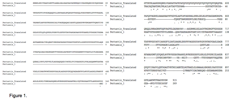
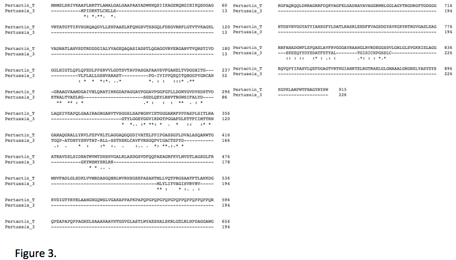
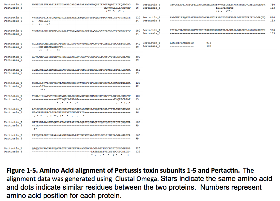
Pertactin and Pertussis toxin (See Below) are both virulence factors that contribute to respiratory tract infection and whooping cough. Both are responsible for binding the foreign bacterial cell to the host organism’s cells. Despite similar functions, Pertactin and Pertussis toxin have very different structures. Pertussis toxin is a virulence factor only produced by Bordetella pertussis. It is known to cause systemic symptoms of pertussis disease, such as leukocytosis and histamine sensitivity. It has also, recently, been discovered to promote respiratory infection by inhibiting and modulating host cell immune responses. Pertussis toxin promotes infection by acting as a soluble factor, which attacks resident cells of the trachea and lungs, such as macrophages[12]. Pertactin is also a virulence factor known to contribute to whooping cough. Pertactin is found in Bordetella pampertussis, and a 91.3% homologous protein is found in Bordetella pertussis, the species which produces Pertussis toxin. An amino acid alignment between the two strains reveals that the proteins differ in the number of repeated sequences. B. pampertussis has a series of approximately twenty more amino acids beginning at the 580th position ( Figure 6). Similarly to Pertussis toxin, Pertactin is involved in binding the bacterial cell to respiratory host cells, however, less is known about the specific cells Pertactin binds to in order to promote infection.[2]
Despite similar functions, Pertussis toxin and Pertactin have very different structures. As mentioned previously, Pertactin’s structure consists of a 16-stranded parallel beta-helix with a v-shaped cross section, while Pertussis toxin consists of five subunits.[2]BLAST results of Pertactin revealed that two domains, autotransporter and PL1_Passenger_AT, are conserved. The autotransporter domain is located at the C-terminus of Pertactin and functions to transport the passenger domain, located at the N-terminus of Pertactin, in to the host cell, where the two domains are typically cleaved.[2] This process is responsible for the bacterial cell binding to the host cell. While Pertactin is a single polypeptide chain, Pertussis toxin consists of five different subunits: S1 makes up subunit A and subunit B is a pentameric ring made of S2, S3, two S4 and S5. BLAST results revealed that a single domain is contained within subunits 1, 4 and 5: Pertussis_S1 superfamily, Pertussis_S4 superfamily and Pertussis_S5 superfamily. Subunits 2 and 3 contain an ATP superfamily and Pertussis_S2S3 Superfamily, which represent the N-terminal domain of aerolysin and pertussis toxin and the C-terminal domain, respectively. The individual structures may be responsible for Pertussis toxin’s ability to recognize receptors on numerous cell types[12].
As a whole, Pertactin and Pertussis toxin are structurally very different. However, amino acid alignments between Pertactin and each of the five subunits of Pertussis toxin reveals that various sections of Pertactin are the same or highly similar to individual subunits of Pertussis toxin ( Figure 1, Figure 2, Figure 3, Figure 4, Figure 5). BLAST results also revealed that subunits 4 and 5’s structures consist of an OB fold and a closed or partly open beta barrel.[2] Similar amino acid sequences, as well as similarities between Pertactin’s beta barrel structure and the beta barrel structures of subunits 4 and 5 may be how Pertactin and Pertussis toxin have similar functions.


Pertussis Toxin
| |||||||||||

3D structures of Pertussis toxin
References
- ↑ 1.00 1.01 1.02 1.03 1.04 1.05 1.06 1.07 1.08 1.09 1.10 1.11 1.12 Hazes B, Boodhoo A, Cockle SA, Read RJ. Crystal structure of the pertussis toxin-ATP complex: a molecular sensor. J Mol Biol. 1996 May 17;258(4):661-71. PMID:8637000 doi:10.1006/jmbi.1996.0277
- ↑ 2.0 2.1 Carbonetti NH, Artamonova GV, Mays RM, Worthington ZE. Pertussis toxin plays an early role in respiratory tract colonization by Bordetella pertussis. Infect Immun. 2003 Nov;71(11):6358-66. PMID:14573656
- ↑ Cornia PB, Hersh AL, Lipsky BA, Newman TB, Gonzales R. Does this coughing adolescent or adult patient have pertussis? JAMA. 2010 Aug 25;304(8):890-6. PMID:20736473 doi:10.1001/jama.2010.1181
- ↑ Bettiol S, Thompson MJ, Roberts NW, Perera R, Heneghan CJ, Harnden A. Symptomatic treatment of the cough in whooping cough. Cochrane Database Syst Rev. 2010 Jan 20;(1):CD003257. PMID:20091541 doi:10.1002/14651858.CD003257.pub3
- ↑ Weiss AA, Johnson FD, Burns DL. Molecular characterization of an operon required for pertussis toxin secretion. Proc Natl Acad Sci U S A. 1993 Apr 1;90(7):2970-4. PMID:8464913
- ↑ 6.0 6.1 Kaslow HR, Burns DL. Pertussis toxin and target eukaryotic cells: binding, entry, and activation. FASEB J. 1992 Jun;6(9):2684-90. PMID:1612292
- ↑ Kenneth Todar, PhD. (2008). http://www.textbookofbacteriology.net/pertussis.html
- ↑ Plaut RD, Carbonetti NH. Retrograde transport of pertussis toxin in the mammalian cell. Cell Microbiol. 2008 May;10(5):1130-9. Epub 2007 Dec 31. PMID:18201245 doi:10.1111/j.1462-5822.2007.01115.x
- ↑ Kenneth Todar, PhD. (2008). http://www.textbookofbacteriology.net/pertussis.html
- ↑ Kenneth Todar, PhD. (2008). http://www.textbookofbacteriology.net/pertussis.html
- ↑ Centers for Disease Control and Prevention. http://www.cdc.gov/pertussis/clinical/treatment.html
- ↑ Altunaiji S, Kukuruzovic R, Curtis N, Massie J. Antibiotics for whooping cough (pertussis). Cochrane Database Syst Rev. 2007 Jul 18;(3):CD004404. PMID:17636756 doi:10.1002/14651858.CD004404.pub3
- ↑ Kenneth Todar, PhD. (2008). http://www.textbookofbacteriology.net/pertussis.html
Supplemental Information
- REDIRECT 1dab
References
- ↑ 1.0 1.1 Benz, I., & Schmidt, M. (n.d.). Structures and functions of autotransporter proteins in microbial pathogens. International Journal of Medical Microbiology, 461-468
- ↑ 2.0 2.1 2.2 2.3 2.4 "Conserved Protein Domain Family." CDD: Conserved Domain Database. NCBI, n.d. Web. 12 Nov. 2015.
- ↑ 3.0 3.1 3.2 3.3 Emsley, P., Charles, I., Fairweather, N., & Isaacs, N. (1996). Structure of Bordetella pertussis virulence factor P.69 pertactin. Nature, 90-92.
- ↑ D'Souza, S. E., Ginsberg, M. H., & Plow, E. F. (1991). Arginyl-glycyl-aspartic acid (RGD): a cell adhesion motif. Trends In Biochemical Sciences, 16(7), 246-250.
- ↑ Williamson, M. P. (1994). The structure and function of proline-rich regions in proteins. Biochemical Journal, 297(Pt 2), 249–260.
- ↑ Gallant, N. D., Michael, K. E., & García, A. J. (2005). Cell Adhesion Strengthening: Contributions of Adhesive Area, Integrin Binding, and Focal Adhesion Assembly. Molecular Biology of the Cell, 16(9), 4329–4340.
- ↑ 7.0 7.1 Henderson, I., & Nataro, J. (2001). Virulence Functions of Autotransporter Proteins. Infection and Immunity, 1231-1243.
- ↑ Junker, M., Schuster, C. C., McDonnell, A. V., Sorg, K. A., Finn, M. C., Berger, B., & Clark, P. L. (2006). Pertactin β-helix folding mechanism suggests common themes for the secretion and folding of autotransporter proteins. Proceedings of the National Academy of Sciences of the United States of America, 103(13), 4918–4923.
- ↑ Charles, I. G., Dougan, G., Pickard, D., Chatfield, S., Smith, M., Novotny, P., … Fairweather, N. F. (1989). Molecular cloning and characterization of protective outer membrane protein P.69 from Bordetella pertussis. Proceedings of the National Academy of Sciences of the United States of America, 86(10), 3554–3558.
- ↑ Gotto, J. W., Eckhardt, T., Reilly, P. A., Scott, J. V., Cowell, J. L., Metcalf, T. N., … Siegel, M. (1993). Biochemical and immunological properties of two forms of pertactin, the 69,000-molecular-weight outer membrane protein of Bordetella pertussis. Infection and Immunity, 61(5), 2211–2215.
- ↑ King A, Berbers G, van Oirschot H, Hoogerhout P, Knipping K, Mooi F (2001). Microbiology 147(11):2885-2895 doi:10.1099/00221287-147-11-2885.
- ↑ 12.0 12.1 Carbonetti, Nicholas H. “Pertussis Toxin and Adenylate Cyclase Toxin: Key Virulence Factors of Bordetella Pertussis and Cell Biology Tools.” Future microbiology 5 (2010): 455–469. PMC. Web. 17 Nov. 2015.
Proteopedia Page Contributors and Editors (what is this?)
Claire Gormley, Melissa Gray, Kimberly Eldridge, Michal Harel, Zackary Zayakosky, Rachel Korba
