Sandbox T1SS T2SS
From Proteopedia
|
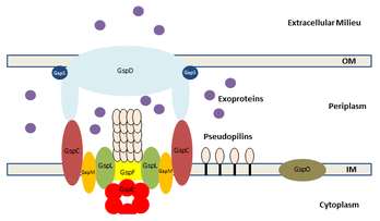
The Type II secretion system (T2SS) is a secretion pathway found in Gram-negative bacteria. Following transport of a protein from the cytoplasm to the periplasm via the Sec or Tat pathway, the T2SS transports proteins from the periplasm to the extracellular environment.[1]
Contents |
Function
Proteins secreted via T2SS include proteases, cellulases, pectinases, phospholipases, lipases, and toxins, many of which are virulence factors.[2] For instance, pathogens such as Vibrio cholerae and Pseudomonas aeruginosa employ the T2SS pathway.[3]
The T2SS is capable of transporting proteins from the periplasm to the extracellular environment. Before moving through the T2SS, a protein must first be transported from the cytoplasm to the periplasm via the Sec or Tat pathway.[4]
Structure

Each T2SS is made of many general secretory proteins (GSPs). Twelve to fifteen GSPs compose one secretory system, and they are often from the same gene cluster and found on the same operon.[4]
The T2SS consists of four core components: an outer membrane complex, an inner membrane complex, an ATPase on the cytoplamsic side, and a pseudopilus. The outer membrane complex consists of the GspD secretin, which uses a beta barrel structure to form a pore through the outer membrane. It is associated with the lipoprotein GspS. The inner membrane complex consists of four proteins: GspC, GspF, GspL, and GspM. Each protein has a different role in secretion, and they all interact with the protein on the periplasmic side. The ATPase is GspE. It uses a Walker A motif to hydrolyze ATP, in order to power the assembly of the pseudopilus, which drives protein secretion. The pseudopilus is composed of GspG, GspH, GspI, GspI and GspK. There is evidence that GspG is the sole component of the stalk portion, which uses a ratcheting motion to push proteins through the membrane.[4]
Energetics
In T2SS, the ATPase GspE hydrolyzes ATP in order to provide the energy for protein secretion. GspE is tightly associated with other inner membrane proteins.[6] GspE hydrolyzes ATP using a Walker A motif, with the sequence GPTGSGKS. It has been suggested that ATP hydrolysis then drives the assembly and disassembly of the pseudopilus, which is composed of GspG. The growth of the pseudopilus is responsible for protein transport, possibly by employing a piston-like motion to push proteins through the channel of the secretion system.[7]
References
- ↑ Saier, Milton H. “Protein Secretion Systems in Gram-Negative Bacteria.” 2006. Microbe 1: 414-419.
- ↑ Sandkvist, Maria. “Type II Secretion and Pathogenesis.” 2001. Infection and Immunity 69: 3523-3535.
- ↑ Douzi B, Filloux A and Voulhoux R (2012). "On the path to uncover the bacterial type II secretion system". Philosophical Transactions of the Royal Society B: Biological Sciences 367: 1059–1072. doi:10.1098/rstb.2011.0204
- ↑ 4.0 4.1 4.2 Cianciotto, Nicholas. “Type II secretion: a protein secretion system for all seasons.” 2005. Trends in Microbiology 13: 581-588.
- ↑ https://en.wikipedia.org/wiki/File:Type_II_secretion_system.png
- ↑ Costa, Tiago, Catarina Felisberto-Rodrigues, Amit Meir, et al. “Secretion systems in Gram-negative bacteria: structural and mechanistic insights.” 2015. Nature Reviews Microbiology 13: 343-359.
- ↑ 10.1074/jbc.M111.234278

