File:OB fold.jpg
From Proteopedia
Jump to navigationJump to search
Size of this preview: 800 × 418 pixels. Other resolution: 2,058 × 1,076 pixels.
Original file (2,058 × 1,076 pixels, file size: 242 KB, MIME type: image/jpeg)
Proteopedia Page Contributors and Editors (what is this?)
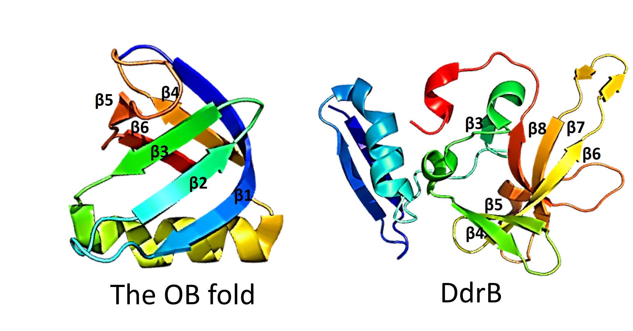
This figure highlights the differences between the classic OB fold found in single-stranded binding proteins and the novel structural features of DdrB. The OB fold is observed in the protein verotoxin-1, PDB code 2XSC . DdrB is modeled from the PDB structure 4HQB. This figure was generated using Pymol.
Proteopedia Page Contributors and Editors (what is this?)
File history
Click on a date/time to view the file as it appeared at that time.
| Date/Time | Thumbnail | Dimensions | User | Comment | |
|---|---|---|---|---|---|
| current | 11:29, 30 April 2014 | | 2,058 × 1,076 (242 KB) | Lauren Ferris (talk | contribs) | This figure highlights the differences between the classic OB fold found in single-stranded binding proteins and the novel structural features of DdrB. The OB fold is observed in the protein verotoxin-1, PDB code 2XSC . DdrB is modeled from the PDB stru |
You cannot overwrite this file.
File usage
The following page uses this file:


