Uploads by Adam Kingsley
From Proteopedia
Jump to navigationJump to search
This special page shows all uploaded files.
| Date | Name | Thumbnail | Size | Description |
|---|---|---|---|---|
| 23:11, 2 May 2022 | GLUT1 Hydrophobic Pocket Sticks 400.png (file) |  |
739 KB | |
| 23:06, 2 May 2022 | GLUT1 Hydrophobic Pocket Sticks.png (file) |  |
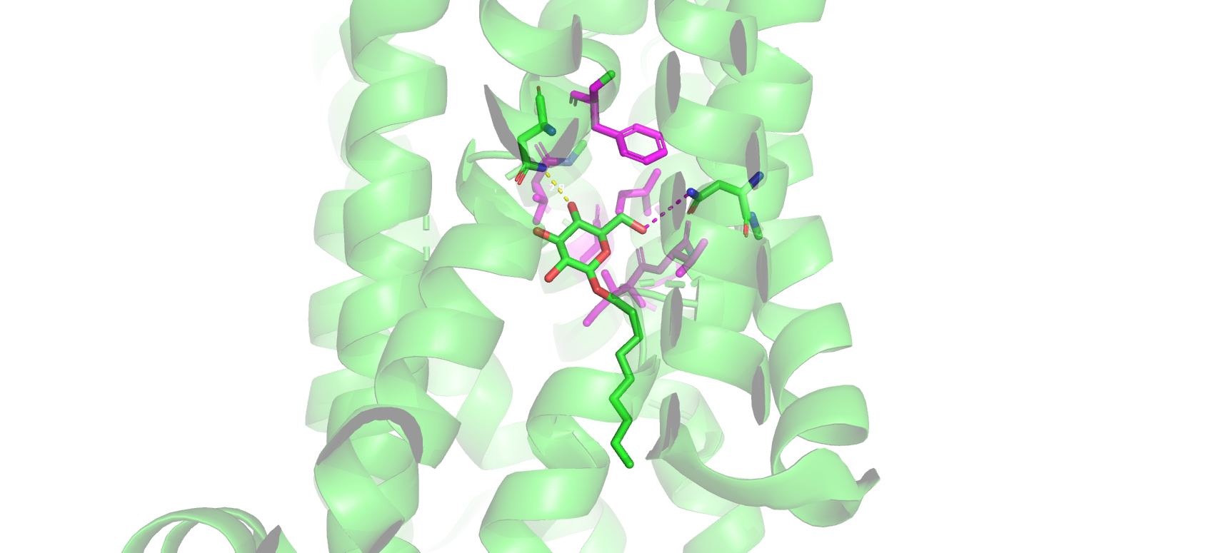
739 KB | An image of the amino acid residues that comprise the glucose-binding site in GLUT1, generated in PyMol. Nonyl β-D-glucopyranoside acts as a glucose analog in the crystal structure 4PYP. |
| 22:48, 2 May 2022 | GLUT1 Hydrophobic Pocket.png (file) |  |
711 KB | Proposed glucose-binding mechanism in the crystal structure 4pyp rendered in PyMol. The proposed hydrophobic pocket residues of Gly27, Thr30, Ile164, Val165, Ile168, and Phe291 are colored magenta. Both Asn288 and Asn317 show that there may also be transi |
| 22:00, 2 May 2022 | GLUT1 Binding Mechanism.png (file) |  |
541 KB | Proposed glucose-binding mechanism in the crystal structure 4pyp rendered in PyMol. The proposed hydrophobic pocket residues of Gly27, Thr30, Ile164, Val165, Ile168, and Phe291 are colored magenta. Both Asn288 and Asn317 show that there may also be tr |
| 00:49, 27 April 2022 | GLUT1 HP Image.png (file) |  |
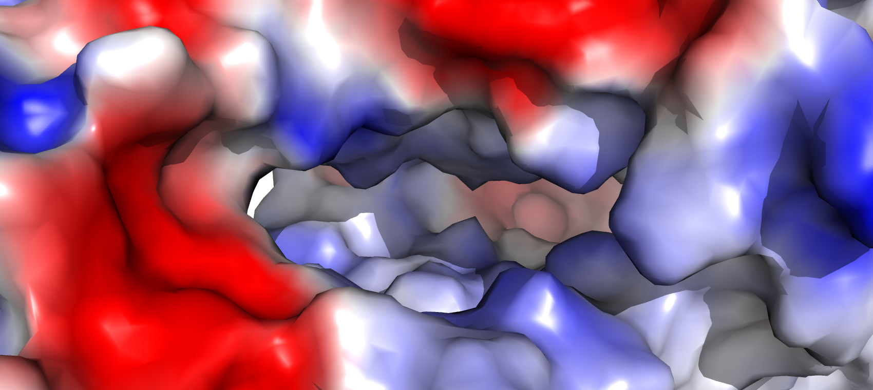
777 KB | Image of the hydrophobic pocket of human GLUT1 generated in PyMol. The surface color indicates the following: red - net negative charge, blue - net positive charge, white - neutral. |
| 20:23, 25 April 2022 | Blood-brain barrier.png (file) |  |
10 KB | This image shows the distribution of different transport proteins in the brain. Astrocytes and capillary endothelial cells express GLUT1. Neurons express GLUT3. Neurons and astrocytes express MCT transporters which transport lactate from astrocytes into n |