Uploads by Ashley R. Wilkinson
From Proteopedia
Jump to navigationJump to search
This special page shows all uploaded files.
| Date | Name | Thumbnail | Size | Description |
|---|---|---|---|---|
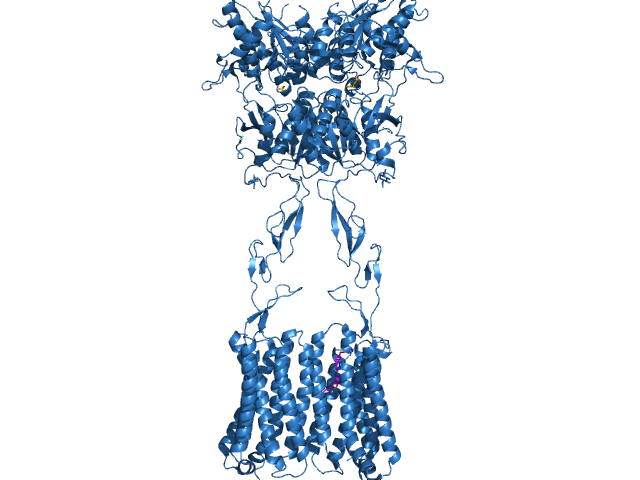
| 17:48, 14 April 2022 | Domains of mGlu2.jpg (file) |  |
123 KB | Extracellular and intracellular domains of mGlu2 |
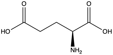
| 14:04, 12 April 2022 | 320px-L-Glutamate Structural Formulae.jpg (file) |  |
9 KB | Glutamate Structure with correct ionization |
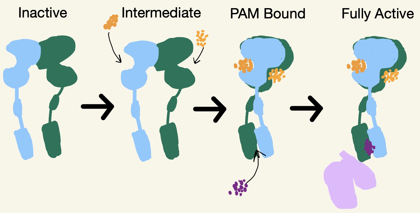
| 13:27, 12 April 2022 | Schematic of mGlu2.jpg (file) |  |
124 KB | States of mGlu2 |
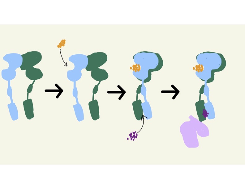
| 13:23, 12 April 2022 | Schematic of mGlu2 States.jpg (file) |  |
53 KB | mGlu2 States |
| 14:51, 29 March 2022 | Morph 7mtq.pdb 7mts.pdb u5090.pdb (file) |  |
12.6 MB | Morph from 7mtq.pdb to 7mts.pdb by Ashley R. Wilkinson Powered by PyMOL from SCHRÖDINGER |
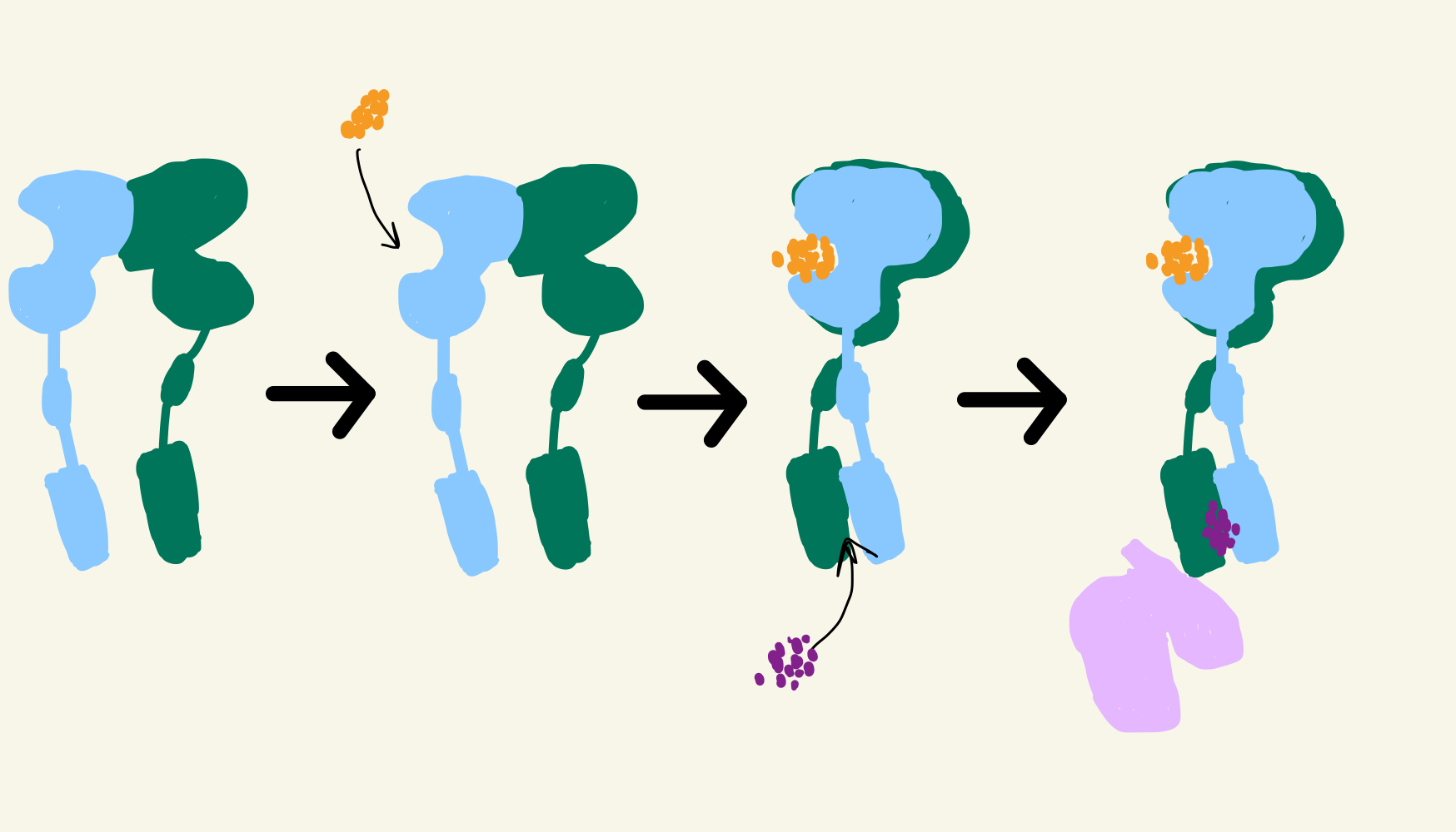
| 14:45, 29 March 2022 | Transformations.jpg (file) |  |
39 KB | |
| 14:44, 29 March 2022 | IMG 1FBB977FEDDF-1.jpeg (file) |  |
293 KB | Transformation of mGlu2 |
| 14:33, 29 March 2022 | IMG 1FBB977FEDDF-1.pdf (file) |  |
297 KB | inactive to fully active schematic |
| 20:21, 28 March 2022 | PAM.pdb (file) | |
867 KB | PAM Bound State |
| 20:18, 28 March 2022 | 7mts.pdb (file) | |
1.24 MB | Active State |
| 20:17, 28 March 2022 | 7mtr.pdb (file) | |
867 KB | PAM Binding Form |
| 19:52, 28 March 2022 | Glutamate.jpeg (file) |  |
12 KB | Structure of Glutamate |
| 14:30, 22 March 2022 | 7mtq.pdb (file) | |
824 KB | This is the inactive structure of mGlu2. |
| 14:13, 22 March 2022 | PAM binding pocket correct.png (file) |  |
94 KB | Figure 3: This is PAM located in its binding pocket. PAM, JNJ-40411813, is shown in magenta and colored by atom. The image shows four labelled alpha helices (III, V, VI, and VII) that create the binding pocket in the 7TM region of mGlu2 for PAM to bind wi |
| 13:17, 22 March 2022 | PAM-1C.png (file) |  |
144 KB |