Uploads by Drew Barber
From Proteopedia
Jump to navigationJump to search
This special page shows all uploaded files.
| Date | Name | Thumbnail | Size | Description |
|---|---|---|---|---|
| 14:21, 30 April 2014 | Reaction of GPx-1.jpg (file) |  |
25 KB | Figure 1. Reaction catalyzed by GPx-1. 1. The peroxidatic Selenocysteine residue reacts with hydrogen peroxide. This yields one molecule of water and the selenenic acid form of the peroxidatic residue (Se-OH). 2. One molecule of glutathione (GSH) react |
| 07:31, 30 April 2014 | Thioredoxin fold.jpg (file) |  |
36 KB | Figure 4. Thioredoxin Fold. Red arrows represent B-sheets while blue rectangles represent alpha-helices. A. Thioredoxin fold. B. Fold of glutathione peroxidase. |
| 06:43, 30 April 2014 | Thioredoxin like fold.jpg (file) |  |
25 KB | Figure 5. Thioredoxin lik fold of GPx-1. Purple sequences are n-term to thioredoxin like fold. Blue sequences represent classic thioredoxin architecture. Red sequences are secondary structures insertions in the thioredoxin fold. |
| 05:32, 30 April 2014 | Helix310.jpg (file) |  |
48 KB | Figure 2. A 3 10 helix from GPx-1 (PDB id: 2F8A) |
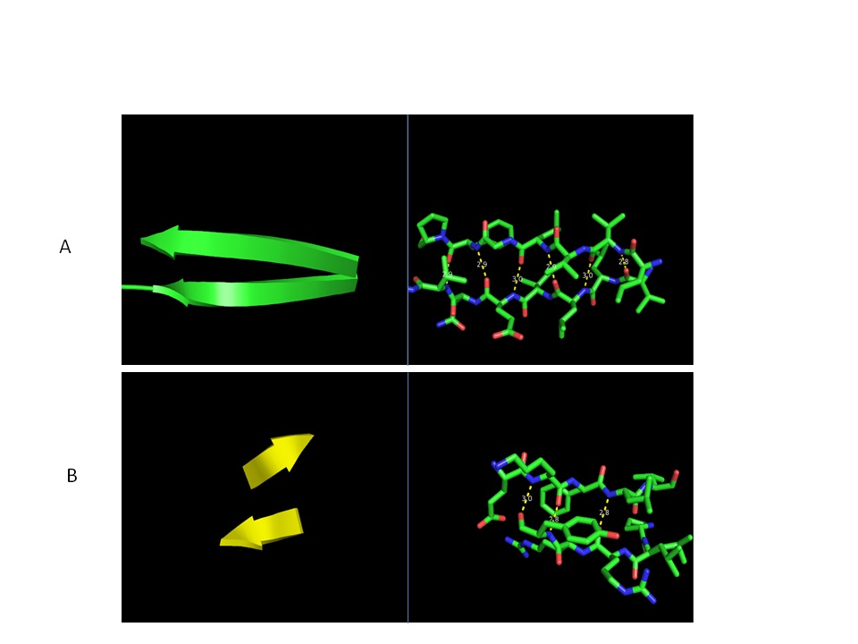
| 05:01, 30 April 2014 | Beta sheets3.jpg (file) |  |
62 KB | Figure 3. Parallel and anti-parallel beta-sheets. A. Parallel beta-sheets represented as cartoon or sticks. Hydrogen bonding is indicated by dashed lines. B. Anti-parallel beta-sheets represented as cartoon or sticks. Hydrogen bonding is indicated by |