Uploads by Natalie Van Ochten
From Proteopedia
Jump to navigationJump to search
This special page shows all uploaded files.
| Date | Name | Thumbnail | Size | Description |
|---|---|---|---|---|
| 18:47, 21 April 2017 | WebLogo for Lid Region.png (file) |  |
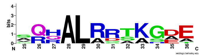
15 KB | WebLogo for the lid region (amino acids 25-36) of DDAH. Sequences come from eleven different organisms to show a clear conservation of a Leucine residue in the lid. Organisms used in this logo include ''Bos taurus'' (cattle), ''Capra hircus'' (goat), ''Ov |
| 18:43, 21 April 2017 | WebLogo for Lid Region .png (file) |  |
15 KB | WebLogo for the lid region (amino acids 25-36) of DDAH. Sequences come from eleven different organisms to show a clear conservation of a Leucine residue in the lid. Organisms used in this logo include "Bos taurus" (cattle), "Capra hircus" (goat), "Ovis ar |
| 18:22, 21 April 2017 | Lid Region WebLogo .png (file) |  |
15 KB | WebLogo for the lid region (amino acids 25-36) of DDAH. Sequences come from many different organisms to show a clear conservation of a Leucine residue in the lid. |
| 18:19, 21 April 2017 | Lid Region WebLogo.png (file) |  |
15 KB | WebLogo for the lid region (amino acids 25-36) of DDAH. Sequences come from many different organisms to show a clear conservation of a Leucine residue in the lid. |
| 14:04, 11 April 2017 | DDAH mechanism.jpg (file) |  |
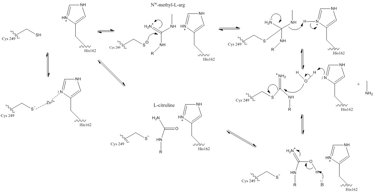
44 KB | The normal DDAH mechanism showing the formation of L-citrulline and mono or dimethylamine from MMA or ADMA. |
| 13:54, 11 April 2017 | DDAH mecahnism.jpg (file) |  |
44 KB | The simple DDAH mecahnism showing the formation of L-citrulline and mono or dimethylamine from MMA or ADMA. |
| 13:45, 11 April 2017 | L-citrulline, L-homocysteine, and S-nitroso-L-homocysteine.jpg (file) |  |
16 KB | Structures of L-citrulline, L-homocysteine, and S-nitroso-L-homocysteine which are DDAH inhibitors that bind to the active site in a similar way. |
| 03:00, 31 March 2017 | Zn(II) bound at differing pH values.jpg (file) | _bound_at_differing_pH_values.jpg) |
280 KB | Zn(II) bound to the active site of DDAH at differing pH values. A) Zn(II) bound at pH 9.0 showing the channel of DDAH. B) Zn(II) bound at 9.0 showing the closed conformation lid with Leu29 blocking the active site. C) Zn(II) bound at pH 6.3 showing the ch |
| 00:41, 31 March 2017 | The Normal DDAH Mechanism.jpg (file) |  |
65 KB | The normal mechanism of DDAH. This figure highlights the importance of Cys249 acting as the nucleophile that attacks the guanidinium carbon on the substrate that is held in the active site via hydrogen bonds. The tetrahedral product collapses as the alkyl |
| 00:31, 31 March 2017 | Proteopediaaaaaaa.jpg (file) |  |
65 KB | The normal mechanism of DDAH. This figure highlights the importance of Cys249 acting as the nucleophile that attacks the guanidinium carbon on the substrate that is held in the active site via hydrogen bonds. The tetrahedral product collapses as the alkyl |
| 13:59, 28 February 2017 | 2CI6 with Zn2+ bound.png (file) |  |
85 KB | This figure shows the crystal form I of Dimethylarginine Dimethylaminohydrolase with Zn2+ bound to the active site at pH 6.3. |