Introduction
Proteinase K can be found on PDB (Protein Data Base) by the ID 2ID8. It is a serine protease encoded by the gene PROK, and cleave other proteins in hydrophobic or aromatic sites.
Proteinase K has a molecular mass of 29373.26 Da [1] and it is a member of the Subtilisin protein family.
Back in 1974, Proteinase K was discovered in extracts of Parengyodontium album (formerly Tritirachium album and Engyodontium album ) and because its ability to digest keratin it received the letter "K". The protein structure was determined from a crystal using X-ray single-wavelength anomalous diffraction (SAD) method in 2006 by Wang et al., but it had previously been determined by Betzel et al. (1988) through X‐ray diffraction at 0.15‐nm resolution.
Proteinase K is largely used in molecular biology protocols to digest proteins and removes contamination from nucleic acid samples, it degrades proteins in cell lysates and deactivates DNAs and RNAses.
Primary Structure
Proteinase K is composite of 279 residues together in only one peptide chain and it was find on the protein surface two calcium ion, tree nitrate anions and one molecule of boric acid, totalizing 2081 atoms (water not included).
The amino acid sequence of Proteinase K is displayed below:
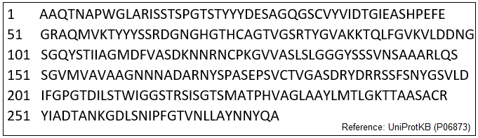
Legend: The amino acid sequence of Proteinase K.
Secundary Structure
Proteinase K secondary structure comprises many alpha-helices and beta-sheets.
- Beginning on the N-terminal, the sequence of these structures is as it follows: BI1-A1-BI2-BII1-BII2-A2-BII3-A3-BII4-(BIII1)-A4-BII5-BII6-BIV1-BIV2-BII7-(BV1-BV2)-A5-A6-BII8-BII9 [2]
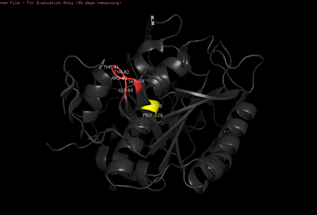
- On alpha-helix 2, the C-terminal is interrupted by an extra loop, that is formed from Ser79 to Gly83 (red in the image below). This is common on other proteases of the subtilisin family, in which there are about five more amino acids in this extra loop. On the alpha-helix 4, there is an incorporation of Pro on the position 228 (Pro228, yellow in the image below). Pro is considered a strong alpha-helix breaker [3].
Those features are shown in the simulation below done in the PyMOL software.
Tertiary Structure
Chemical bonds
Disulfide Bridges
Although Proteinase K presents five cysteine amino acids, only four of them participates in disulfide bridges.
There are show two disulfide bridges on this protein. These bridges are made by the presence of cysteines on the central beta-sheet, on the positions 34 and 178 (Cys34 and Cys178). One disulfide bridge is the Cys34-Cys123 interactions, and the other is Cys178-Cys249 [4]. The fifth Cysteine 73 is not participating in any disulfide bridge.
Hidrogen Bonds
The participating amino acids in hydrogen bonding between peptides include 182 out of the total 279 (or 65.2%) [5].
The folding of the protein is stabilized by a total of 130 hydrogen bonds, of which 55 are made between the main chain and side chain functional groups, and 75 are made between side chains. If we include the water molecules, the hydrogen bonds interactions rise up to 191 interactions. [6].
Binding sites
Active Site and Substrate Recognition
The catalytic site of Proteinase K is made by a triad of amino acids, Asp39, His69 and Ser224 [7] (hence it is a serine protease). The pink color indicates that these amino acids are all polars. See more about Proteinase K function in the Function section below.
The amino acids side chains of the catalytic triad are stabilized by hydrogen bonds, as demonstrated by a simulation done in the PyMOL software. We know that these are hydrogen bonds because the distance between the atoms participating in the ligation is around 2.8 angstroms. See the simulation:
 .
.
Media: Atomic_distance_catalytic_site_of_Proteinase_K_.mp4.
Legend: The atomic distance (Angstroms) between the catalytic amino acid triad. Simulation made through PyMOL software.
The active site of the protein is located on the central beta-sheet (as described above). The Asp39 aminoacid accepts two hydrogen bonds, on the carbonyl oxygen, from Val95 and Leu96, in a way that stabilizes this important residue [8].
The susbtrate recognition are made by two peptide chains, one that englobes aminoacids ranging from 99 to 104 positions, and the other made of aminoacids ranging from 132 to 136 positions, which can be viewed here. The colors indicate where there is a region with alpha-helix (in pink), beta-sheet (in gold), or loop (in white).
Calcium binding sites
There are two calcium binding sites in Proteinase K. Calcium ions contribute to the stabilization of the overall structure of the protein.
The amino acids participating in one of the binding sites are Pro175, Val177, and Asp200, as shown here.
he other calcium binding site is less well-defined [9], and are formed by the amino acids Thr16 and Asp260, as shown here. Calcium is not represented in this scene.
Other structural highlights
- The N-terminal and C-terminal are close to each other by interactions made by the Thr16 and Asp260, and by Arg12 and Asp260, Asp187 [10], which can be viewed here.
- Following the structure, the protein folds forming a beta-sheet that has eight parallel strands. This beta-sheet forms the central core of proteinase K, and has five alpha-helices and more three antiparallel beta-sheets, outside this central beta-sheet [11].
Function
Proteinase K possesses the ability to digest a huge variety of proteins. The cleavage occurs preferably on hydrophobic or aromatic amino acids.
As it was already talked above, the catalytic site of Proteinase K is made up of three amino acids. Two of them (His69 and Ser224) are located on the N-terminal of alpha-helices. This is important for the function of the enzyme, because of the polarity. This positive charge may help stabilize the peptide that has been cleaved because once the reaction has occurred, there is a formation of negatively charged oxyanion.
- There are two distinct functions in the active center [12]. One is the catalytic site, described before, which includes the triad of amino acids (Asp39, His69, Ser224) involved in the hydrolysis of a substrate (peptides). The other function is the substrate recognition, made by two peptide chains. Both of these sequences (the tirade and the sequences that recognize the substrate) are conserved in the subtilisin family.
- Calcium is important for the enzymatic function of Proteinase K [13].Only one of the two calcium that binds to the protein is found attached on the protein during protocols for structure analyses, and if this ion is removed, the activity of Proteinase K decreases [14], showing a importance of this ligation for the normal function of the enzyme.
Relevance
Laboratory protocols
- Since Proteinase K digest native proteins, and it is stable in front of high temperatures, urea, and others denaturacting factors, this enzyme is used on the purification on preparations of nucleic acids, and it is also used to digest proteins (that have hydrophobic or aromatic amino acids). Thus, proteinase K is widely used by molecular biology laboratories on protocols on the most diverse types of experiments to ensure the elimination of DNase, RNase, and proteins from its preparations.
Clinic
- Proteinase K was historically used to perform Prion disease diagnostic. The disease-related form of prion protein (PrPSc) presents resistance to proteolysis, hence, the diagnostic of Prion disease rely upon the detection of resistant protein forms to degradation by proteinase [15].
- ↑ PDB 2ID8: Wang, Jiawei, Miroslawa Dauter, and Zbigniew Dauter. "What can be done with a good crystal and an accurate beamline?." Acta Crystallographica Section D: Biological Crystallography 62.12 (2006): 1475-1483. Available: https://www.ncbi.nlm.nih.gov/pubmed/17139083
- ↑ Betzel, Christian, Gour P. PAL, and Wolfram SAENGER. "Three‐dimensional structure of proteinase K at 0.15‐nm resolution." The FEBS Journal 178.1 (1988): 155-171. Available: https://onlinelibrary.wiley.com/doi/abs/10.1111/j.1432-1033.1988.tb14440.x
- ↑ Betzel, Christian, Gour P. PAL, and Wolfram SAENGER. "Three‐dimensional structure of proteinase K at 0.15‐nm resolution." The FEBS Journal 178.1 (1988): 155-171. Available: https://onlinelibrary.wiley.com/doi/abs/10.1111/j.1432-1033.1988.tb14440.x
- ↑ Betzel, Christian, Gour P. PAL, and Wolfram SAENGER. "Three‐dimensional structure of proteinase K at 0.15‐nm resolution." The FEBS Journal 178.1 (1988): 155-171. Available: https://onlinelibrary.wiley.com/doi/abs/10.1111/j.1432-1033.1988.tb14440.x
- ↑ Betzel, Christian, Gour P. PAL, and Wolfram SAENGER. "Three‐dimensional structure of proteinase K at 0.15‐nm resolution." The FEBS Journal 178.1 (1988): 155-171. Available: https://onlinelibrary.wiley.com/doi/abs/10.1111/j.1432-1033.1988.tb14440.x
- ↑ Betzel, Christian, Gour P. PAL, and Wolfram SAENGER. "Three‐dimensional structure of proteinase K at 0.15‐nm resolution." The FEBS Journal 178.1 (1988): 155-171. Available: https://onlinelibrary.wiley.com/doi/abs/10.1111/j.1432-1033.1988.tb14440.x
- ↑ Betzel, Christian, Gour P. PAL, and Wolfram SAENGER. "Three‐dimensional structure of proteinase K at 0.15‐nm resolution." The FEBS Journal 178.1 (1988): 155-171. Available: https://onlinelibrary.wiley.com/doi/abs/10.1111/j.1432-1033.1988.tb14440.x
- ↑ Betzel, Christian, Gour P. PAL, and Wolfram SAENGER. "Three‐dimensional structure of proteinase K at 0.15‐nm resolution." The FEBS Journal 178.1 (1988): 155-171. Available: https://onlinelibrary.wiley.com/doi/abs/10.1111/j.1432-1033.1988.tb14440.x
- ↑ Betzel, Christian, Gour P. PAL, and Wolfram SAENGER. "Three‐dimensional structure of proteinase K at 0.15‐nm resolution." The FEBS Journal 178.1 (1988): 155-171. Available: https://onlinelibrary.wiley.com/doi/abs/10.1111/j.1432-1033.1988.tb14440.x
- ↑ Betzel, Christian, Gour P. PAL, and Wolfram SAENGER. "Three‐dimensional structure of proteinase K at 0.15‐nm resolution." The FEBS Journal 178.1 (1988): 155-171. Available: https://onlinelibrary.wiley.com/doi/abs/10.1111/j.1432-1033.1988.tb14440.x
- ↑ Betzel, Christian, Gour P. PAL, and Wolfram SAENGER. "Three‐dimensional structure of proteinase K at 0.15‐nm resolution." The FEBS Journal 178.1 (1988): 155-171. Available: https://onlinelibrary.wiley.com/doi/abs/10.1111/j.1432-1033.1988.tb14440.x
- ↑ Betzel, Christian, Gour P. PAL, and Wolfram SAENGER. "Three‐dimensional structure of proteinase K at 0.15‐nm resolution." The FEBS Journal 178.1 (1988): 155-171. Available: https://onlinelibrary.wiley.com/doi/abs/10.1111/j.1432-1033.1988.tb14440.x
- ↑ Betzel, Christian, Gour P. PAL, and Wolfram SAENGER. "Three‐dimensional structure of proteinase K at 0.15‐nm resolution." The FEBS Journal 178.1 (1988): 155-171. Available: https://onlinelibrary.wiley.com/doi/abs/10.1111/j.1432-1033.1988.tb14440.x
- ↑ Betzel, Christian, Gour P. PAL, and Wolfram SAENGER. "Three‐dimensional structure of proteinase K at 0.15‐nm resolution." The FEBS Journal 178.1 (1988): 155-171. Available: https://onlinelibrary.wiley.com/doi/abs/10.1111/j.1432-1033.1988.tb14440.x
- ↑ Cronier, Sabrina, et al. "Detection and characterization of proteinase K-sensitive disease-related prion protein with thermolysin." Biochemical Journal 416.2 (2008): 297-305. Available online: https://www.biochemj.org/content/416/2/297


