3ad8
From Proteopedia
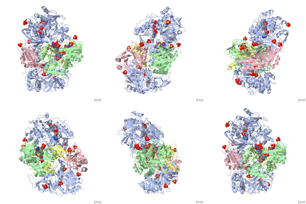
Heterotetrameric Sarcosine Oxidase from Corynebacterium sp. U-96 in complex with pyrrole 2-carboxylate
Structural highlights
FunctionEvolutionary ConservationCheck, as determined by ConSurfDB. You may read the explanation of the method and the full data available from ConSurf. Publication Abstract from PubMedWe characterized the crystal structures of heterotetrameric sarcosine oxidase (SO) from Corynebacterium sp. U-96 complexed with methylthioacetate (MTA), pyrrole 2-carboxylate (PCA), and sulfite, and of sarcosine-reduced SO. SO comprises alpha, beta, gamma , and delta subunits; FAD and FMN cofactors; and a large internal cavity. MTA and PCA are sandwiched between the re-face of the FAD isoalloxazine ring and the beta subunit C-terminal residues. Reduction of flavin cofactors shifts the beta subunit Ala1 toward the alpha subunit Met55, forming a surface cavity at the oxygen-channel vestibule and rendering the beta subunit C-terminal residues mobile. We identified three channels connecting the cavity and the enzyme surface. Two of them exist in the intersubunit space between alpha and beta subunits, and the substrate sarcosine seems to enter the active site through either of these channels and reaches the re-side of the FAD isoalloxazine ring by traversing the mobile beta subunit C-terminal residues. The third channel goes through the alpha subunit and has a folinic acid-binding site, where the iminium intermediate is converted to Gly and either formaldehyde or, 5,10-methylenetetrahydrofolate. Oxygen molecules are probably located on the surface cavity and diffuse to the FMN isoalloxazine ring; the H(2)O(2) formed exits via the oxygen channel. Channeling and conformational changes in the heterotetrameric sarcosine oxidase from Corynebacterium sp. U-96.,Moriguchi T, Ida K, Hikima T, Ueno G, Yamamoto M, Suzuki H J Biochem. 2010 Aug 18. PMID:20675294[1] From MEDLINE®/PubMed®, a database of the U.S. National Library of Medicine. See AlsoReferences
| ||||||||||||||||||||


