Sphingosine kinase
From Proteopedia
| |||||||||||
Contents |
Sphingosine Kinases are Evolutionarily Conserved
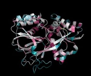
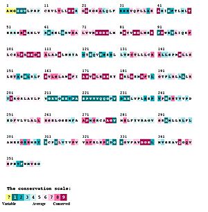
Sphingosine Kinases are part of distinct class of lipid kinases that is evolutionarily highly conserved (Figure 2 and 3).[2] They do not share sequence homology with other lipid kinases, such as phosphotidylinositol-3 kinase (PI3K)(Figure 3). [1] However, protein sequence motif analysis and structure classification suggested that SphKs belong to the phosphofructokinase (PFK)-like superfamily [3] [4] sharing the same protein fold (i.e. the Rossmann fold) with NAD kinases, ceramide kinases, and phosphofructokinases. [1]
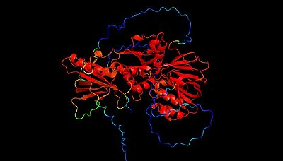
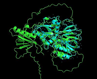
The mammalian isoforms of sphingosine kinase, SphK1 and SphK2, are the only isoforms that have been characterized (Figure 4 and 5). Additionally, the crystal structure has been solved for SphK1 (via X-ray diffraction resulting in a resolution of 2.00Å) with SphK2 having a highly predictive Alpha Fold structure (Figure 4 and 5). [1] Figure 5 shows the alignment of the crystal structure of SphK1 and the Alpha Fold prediction model of SphK2. Here, the catalytic domain aligns with high confidence (showing an RMSD value of 0.0846Å. However, the Alpha Fold prediction model of Sphk2 shows an entire domain (of α/β structure architecture) that does not align with the SphK1 crystal structure. As you will read further in the "Sphingosine Kinase (SphK1 and SphK2)" section, although SphK1 and SphK2 share similar structural properties and mechanistic properties, their kinetic regulation is different (a reason as to why there may be an additional domain predicted in the SphK2 structure).
Sphingolipids
Sphingolipids are a class of lipids, containing a sphingosine base moiety, essential for eukaryotic cell membrane structure and function. [5] Importantly, sphingolipids can additionally act as critical signaling molecules used in many eukaryotic homeostatic cellular processes, such as inflammation, proliferation, apoptosis, cell migration, and pathogen defense. [5] Because of this, it is critical for sphingolipids and their metabolizing enzymes to exist in a delicate homeostatic balance within the eukaryotic cell system.
Sphingosine-1-Phosphate (S1P), a lipid within the sphingolipid class, is an important signaling molecule that can participate in intracellular and extracellular signaling. [5] [6] If meant to be an intracellular signaling molecule, S1P is important for cell survival, growth, and overall cell function.[6] Alternatively, if S1P is meant to be an extracellular signaling molecule, later binding to one of five different S1P G-Protein Coupled Receptors on the same or different cell type, it is used to activate many signaling cascades important for cellular response. [6] One important function of S1P, whether its fate serves intracellularly or extracellularly, is eliciting the immune response (i.e. lymphocyte trafficking), especially as a mechanism in pathogen defense (please see section below labeled "Importance of SphKs in Pathogen Defense and Immune Response" for more information about SphKs involvement within the immune response). [6]
Sphingosine Kinase (SphK1 and SphK2)
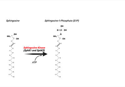
Sphingosine Kinase, existing in two isoenzyme forms, SphK1 and SphK2, creates S1P via the phosphorylation of sphingosine, the base moiety of all sphingolipids (Figure 6). [6] Particularly, this catalytic reaction is ATP (adenine triphosphate)-dependent aiding the phosphorylation of sphingosine on the primary hydroxyl group.
Sphingosine Kinase(s) are a key enzyme controlling the levels of S1P within the eukaryotic cell, and thus, is an important regulator of diverse cellular functions (as mentioned previously). [1] As there aretwo isoforms of sphingosine kinase, SphK1 and SphK2, each isoform has it own specificities. [7] For instance, SphK1 and SphK2 are tissue specific. SphK1 is prevalent in the lung and spleen, whereas SphK2 is prevalent in the liver and heart. [6] Importantly, although SphK1 and SphK2 catalyze the same biochemical reactions, they originate from different genes and have different substrate specificities, tissue distributions (like mentioned above), and subcellular localization patterns.[1] Typically sphingolipid metabolizing enzymes are located along the Endoplasmic Reticulum (ER). [6] Particularly, SphK2 has 4 transmembrane domains and is thus localized to the membrane of the ER where it serves to phosphorylate circulating sphingosine when signaled to do so. [6] Alternatively, SphK1 has no transmembrane domains (being, instead, located within the cytoplasm close to the ER organelle). [6] However, it is unknown whether SphK1 is tethered by some other molecule to the ER or remains untethered within the cytoplasmic compartment. Although SphK1 and SphK2 are similar in function, they are additionally diverse in the localization within the eukaryotic cell environment with differing kinetic properties (i.e., prevalent kinetic regulation differences). [6]
Importance of SphKs in Pathogen Defense and Immune Response
Upon the SphK ATP-dependent phosphorylation event of sphingosine to S1P, the phosphorylated lipid product can be utilized in many intracellular and extracellular signaling mechanisms. [7] One critical function of S1P (through autocrine or paracrine extracellular signaling), is eliciting the immune response when pathogens are present or causing infection. [8] In order to "kick-start" the immune response, first, S1P is transported out of the cell by Spinster Homologue 2, SPNS2, a member of a large family of non ATP-dependent ion transporters. [9] After the lipid's transport out of eukaryotic cell, S1P can bind to one of 5 S1P G-protein coupled receptors (GPCRs) present on the surface of both innate and adaptive immune cell types resulting in immune cell trafficking to the site of infection. [9] [8] For example, macrophages express S1P receptors I, II, III, and IV. Upon binding of S1P to S1P receptor type I, macrophages are signaled to be trafficked and recruited to the site of pathogen infection. [8]
3D structures of sphingosine kinase
Updated on 19-January-2023
4v24 – hSphK1 kinase domain 1-363 + pyrrolidine derivative – human
3vzb – hSphK1 kinase domain + sphingosine
3vzc, 4l02 – hSphK1 kinase domain + inhibitor
3vzd – hSphK1 kinase domain + inhibitor + ADP
References
- ↑ 1.00 1.01 1.02 1.03 1.04 1.05 1.06 1.07 1.08 1.09 1.10 1.11 Wang Z, Min X, Xiao SH, Johnstone S, Romanow W, Meininger D, Xu H, Liu J, Dai J, An S, Thibault S, Walker N. Molecular Basis of Sphingosine Kinase 1 Substrate Recognition and Catalysis. Structure. 2013 Apr 16. pii: S0969-2126(13)00086-5. doi:, 10.1016/j.str.2013.02.025. PMID:23602659 doi:http://dx.doi.org/10.1016/j.str.2013.02.025
- ↑ Kohama T, Olivera A, Edsall L, Nagiec MM, Dickson R, Spiegel S. Molecular cloning and functional characterization of murine sphingosine kinase. J Biol Chem. 1998 Sep 11;273(37):23722-8. PMID:9726979
- ↑ Cheek S, Ginalski K, Zhang H, Grishin NV. A comprehensive update of the sequence and structure classification of kinases. BMC Struct Biol. 2005 Mar 16;5:6. doi: 10.1186/1472-6807-5-6. PMID:15771780 doi:http://dx.doi.org/10.1186/1472-6807-5-6
- ↑ Labesse G, Douguet D, Assairi L, Gilles AM. Diacylglyceride kinases, sphingosine kinases and NAD kinases: distant relatives of 6-phosphofructokinases. Trends Biochem Sci. 2002 Jun;27(6):273-5. doi: 10.1016/s0968-0004(02)02093-5. PMID:12069781 doi:http://dx.doi.org/10.1016/s0968-0004(02)02093-5
- ↑ 5.0 5.1 5.2 Futerman AH, Hannun YA. The complex life of simple sphingolipids. EMBO Rep. 2004 Aug;5(8):777-82. doi: 10.1038/sj.embor.7400208. PMID:15289826 doi:http://dx.doi.org/10.1038/sj.embor.7400208
- ↑ 6.0 6.1 6.2 6.3 6.4 6.5 6.6 6.7 6.8 6.9 Spiegel S, Milstien S. Sphingosine-1-phosphate: an enigmatic signalling lipid. Nat Rev Mol Cell Biol. 2003 May;4(5):397-407. doi: 10.1038/nrm1103. PMID:12728273 doi:http://dx.doi.org/10.1038/nrm1103
- ↑ 7.0 7.1 Futerman AH, Hannun YA. The complex life of simple sphingolipids. EMBO Rep. 2004 Aug;5(8):777-82. doi: 10.1038/sj.embor.7400208. PMID:15289826 doi:http://dx.doi.org/10.1038/sj.embor.7400208
- ↑ 8.0 8.1 8.2 Futerman AH, Hannun YA. The complex life of simple sphingolipids. EMBO Rep. 2004 Aug;5(8):777-82. doi: 10.1038/sj.embor.7400208. PMID:15289826 doi:http://dx.doi.org/10.1038/sj.embor.7400208
- ↑ 9.0 9.1 Spiegel S, Maczis MA, Maceyka M, Milstien S. New insights into functions of the sphingosine-1-phosphate transporter SPNS2. J Lipid Res. 2019 Mar;60(3):484-489. doi: 10.1194/jlr.S091959. Epub 2019 Jan 17. PMID:30655317 doi:http://dx.doi.org/10.1194/jlr.S091959
