Terminal Uridylyl Transferase
From Proteopedia
Contents |
Uridylyl transferases
INTRODUCTION
Terminal uridylyl transferases (TUTases) belong to a superfamily of polymerase ß nucleotidyl transferases.[1] TUTases have been isolated from Trypanosoma brucei and also Leishmania ssp, parasites that cause diseases in humans such as African Sleeping Sickness. It has been suggested that the knowledge of TUTases may aid in the treatment of these diseases as TUTases function in RNA editing in these parasites, and thus can serve as enzymes to target.[2] More specifically TUT4 catalyzes a reaction that adds a nucleotide, from a nucleotide triphosphate, to uridine monophosphate (UMP), the minimally required terminal RNA substrate.[1] TUTase4 is able to bind to the nucleotide triphosphates ATP, CTP, GTP or UTP, however, UTP and CTP are preferred, whereas ATP and GTP ligands have been shown to cause a significant decrease in enzymatic activity.[1] The preference for UTP causes TUTase4 to typically add a uracil nucleotide to the RNA substrate. This selectivity has a variety of mechanisms, including a loss of coplanarity (π-electron stacking) between the ATP and a tyrosine of the active site (Y189) required for catalysis, and reduced stacking between the UMP and ATP rings.[1] The RNA substrate in trypanosomal TUTases selects for cognate nucleosides and provides a metal ion binding site for Mg2+ ions required by the ligand.[1]
| |||||||||
| 2q0d, resolution 2.00Å () | |||||||||
|---|---|---|---|---|---|---|---|---|---|
| Ligands: | , | ||||||||
| Gene: | TUT4 (Trypanosoma brucei) | ||||||||
| Activity: | RNA uridylyltransferase, with EC number 2.7.7.52 | ||||||||
| Related: | 2ikf, 2nom | ||||||||
| |||||||||
| |||||||||
| Resources: | FirstGlance, OCA, RCSB, PDBsum | ||||||||
| Coordinates: | save as pdb, mmCIF, xml | ||||||||
STRUCTURE
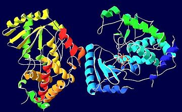
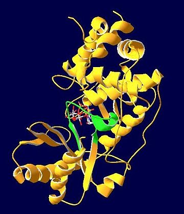
TUT4 with a bound (consisting of an ATP molecule and two Mg2+ ions) has little π-electron stacking with both the active site (Y189) and the RNA substrate, and so is destabilizing, however the phosphate groups of the ATP have been shown to superpose well with that of the other ligands.[1] The Mg2+ ions are coordinated by three (D66, D68, and D136) which are conserved among TUTases, and thus vital in the transferase reaction.[1] Hydrogen bonding and hydrophobic interactions are important in the binding of the RNA substrate to the enzyme as well as the binding of the ligand to the apo protein. Notably, hydrogen bonding interactions occur among of TUT4 with the RNA substrate, and among of the apo protein with the ATP complex.[1] Hydrophobic interactions with the RNA substrate and of TUT4 also contribute to the transferase reaction.[1] The lack of triple stacking as well as different hydrogen bonding interactions contribute to the preference of TUT4 for UTP instead of ATP, however it is thought that minimal mutations would be required for TUT4 to become ATP specific. [1] The signature active site motif for the polymerase β nucleotidyltransferase superfamily, including TUT4 is hG [G/S]X(9-13)Dh[D/E]h (where X designates any amino acid, and h designates hydrophobic amino acids).[2]
TRANSFERASE REACTION
In the most general sense, the transferase reaction consists of the RNA substrate nucleophile (with some nucleotide selectivity) attacking the α-phosphorus atom of the nucleotide triphosphate ligand.[1] The Mg2+ ions are an important component of this reaction as one is thought to aid nucleophile deprotonation with the catalytic base (expected to be D136) and the other is thought to stabilize the leaving group (pyrophosphate).[1] However, due to steric constraints between the ATP ligand and the active site and RNA substrate, RNA binding is destabilized, thus slowing catalysis and the transfer of adenosine nucleotides.[1]
REFERENCES
- ↑ 1.00 1.01 1.02 1.03 1.04 1.05 1.06 1.07 1.08 1.09 1.10 1.11 1.12 Stagno J, Aphasizheva I, Aphasizhev R, Luecke H. Dual role of the RNA substrate in selectivity and catalysis by terminal uridylyl transferases. Proc Natl Acad Sci U S A. 2007 Sep 11;104(37):14634-9. Epub 2007 Sep 4. PMID:17785418
- ↑ 2.0 2.1 Aphasizhev R, Sbicego S, Peris M, Jang SH, Aphasizheva I, Simpson AM, Rivlin A, Simpson L. Trypanosome mitochondrial 3' terminal uridylyl transferase (TUTase): the key enzyme in U-insertion/deletion RNA editing. Cell. 2002 Mar 8;108(5):637-48. PMID:11893335
See Also
2q0d is TUT4 with bound ATP
External Links
RCSB Protein Data Bank: TUT4 with bound ATP
RCSB Protein Data Bank: TUT4 with bound CTP
RCSB Protein Data Bank: TUT4 with bound UTP and UMP


