Uploads by Lauren Ferris
From Proteopedia
Jump to navigationJump to search
This special page shows all uploaded files.
| Date | Name | Thumbnail | Size | Description |
|---|---|---|---|---|
| 12:00, 30 April 2014 | DNA T4-6.jpg (file) |  |
221 KB | Interactions between DdrB and T4-T6. (PDB: 4HQB) This figure was generated using Pymol. |
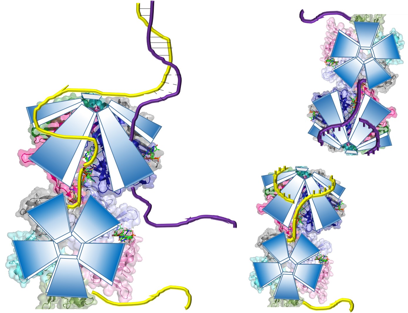
| 11:52, 30 April 2014 | Pentamer-pentamer.jpg (file) |  |
234 KB | Model of the pentamer-pentamer structure. (PDB: 4HQB) This figure was generated using Pymol. |
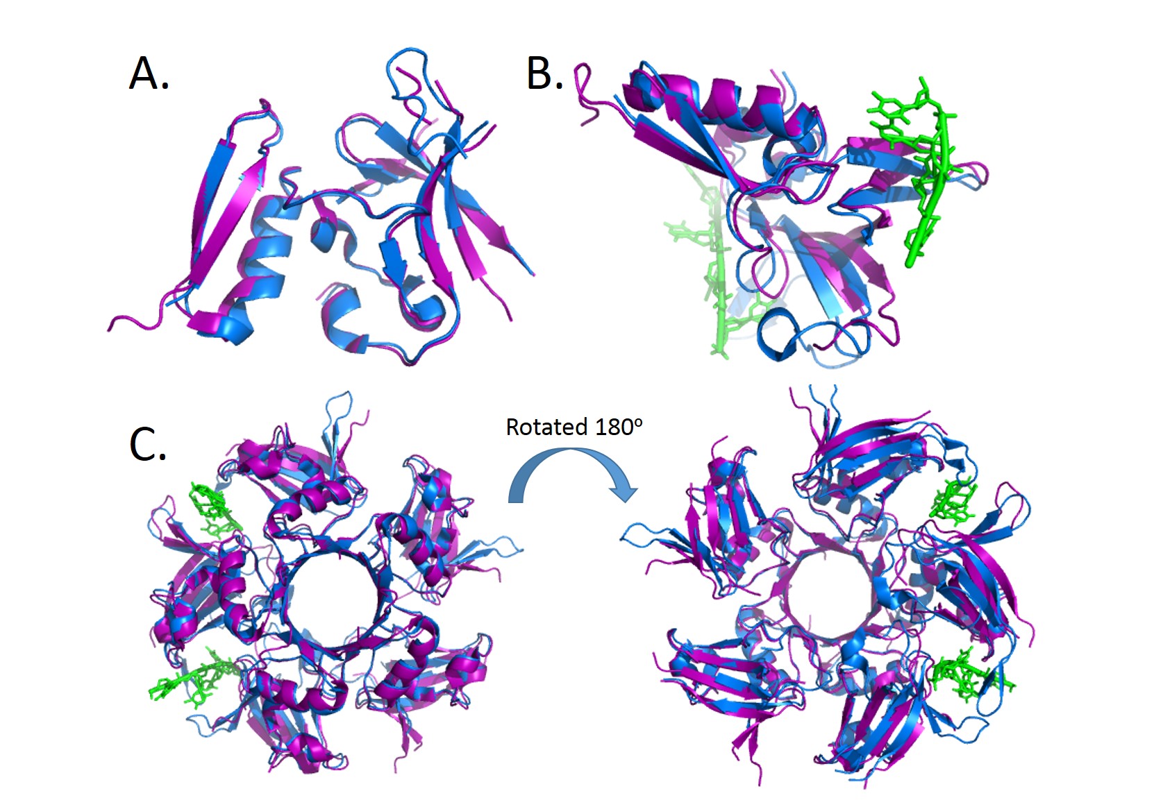
| 11:44, 30 April 2014 | Alignment.jpg (file) |  |
330 KB | Alignment of the crystal structures of Deinococcus radiodurans and Deinococcus geothermalis. (A.) Alignment of chains c, (B.) Alignment of Chains a with DNA, (C.) Alignment of the pentamer. (PDB: 4EXW and 4HQB) This figure was generated using Pymol. |
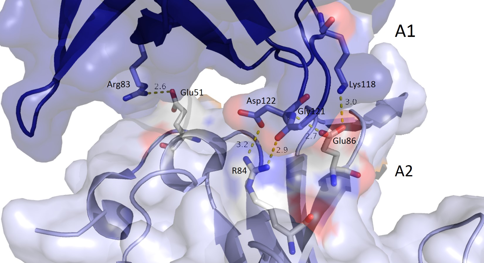
| 11:42, 30 April 2014 | A chains.jpg (file) |  |
191 KB | The pentamer-pentamer interface is also stabilized by salt bridges and hydrogen bonds between the A chainds. (PDB:4HQB) This figure was generated using Pymol. |
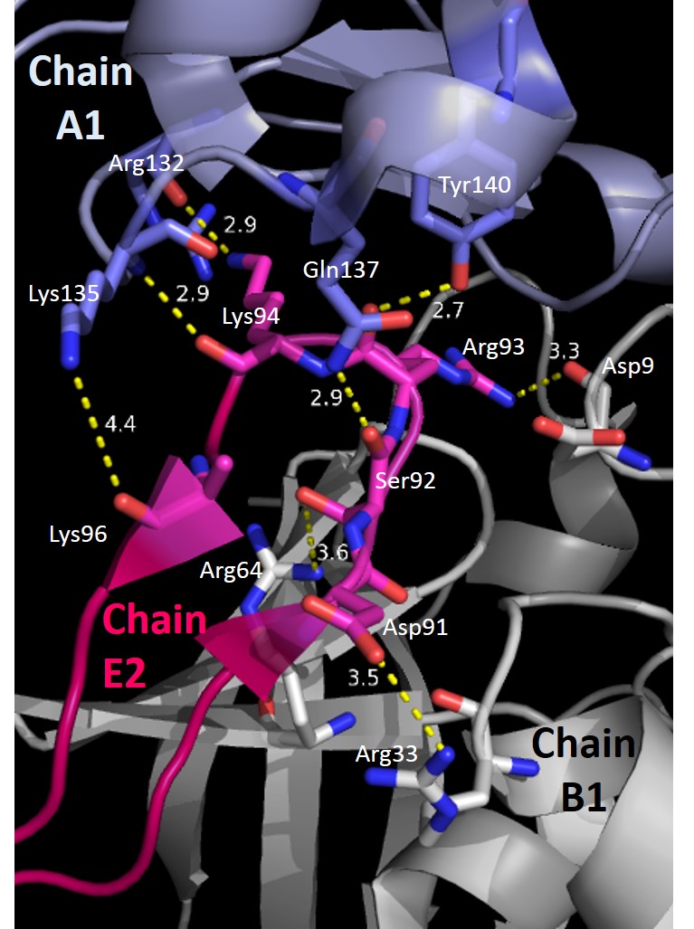
| 11:41, 30 April 2014 | E loop.jpg (file) |  |
134 KB | The pentamer-pentamer interactions are stabilized by interactions involving the hairpin of chain E. (PDB:4HQB). This figure was generated using Pymol. |
| 11:39, 30 April 2014 | T7-8.jpg (file) |  |
294 KB | Interactions between DdrB and T7 and T8. (PDB: 4HQB) This figure was generated using Pymol. |
| 11:35, 30 April 2014 | T1-3.jpg (file) |  |
211 KB | Interactions between DdrB and T1-T3. (PDB: 4HQB) This figure was generated using Pymol. |
| 11:33, 30 April 2014 | Electrostatics pentamer.jpg (file) |  |
163 KB | The electrostatic charges in the DdrB pentamer. Blue represents positive charges, while red represents negative charges. A positive “track” around the top of the pentamer may enable ssDNA binding to one side of the pentamer (left image). (PDB: 4HQB) |
| 11:32, 30 April 2014 | Electrostatics monomer.jpg (file) |  |
177 KB | The electrostatic charges in the DdrB monomer. Blue represents positive charges, while red represents negative charges. (PDB: 4HQB). This figure was generated using Pymol. |
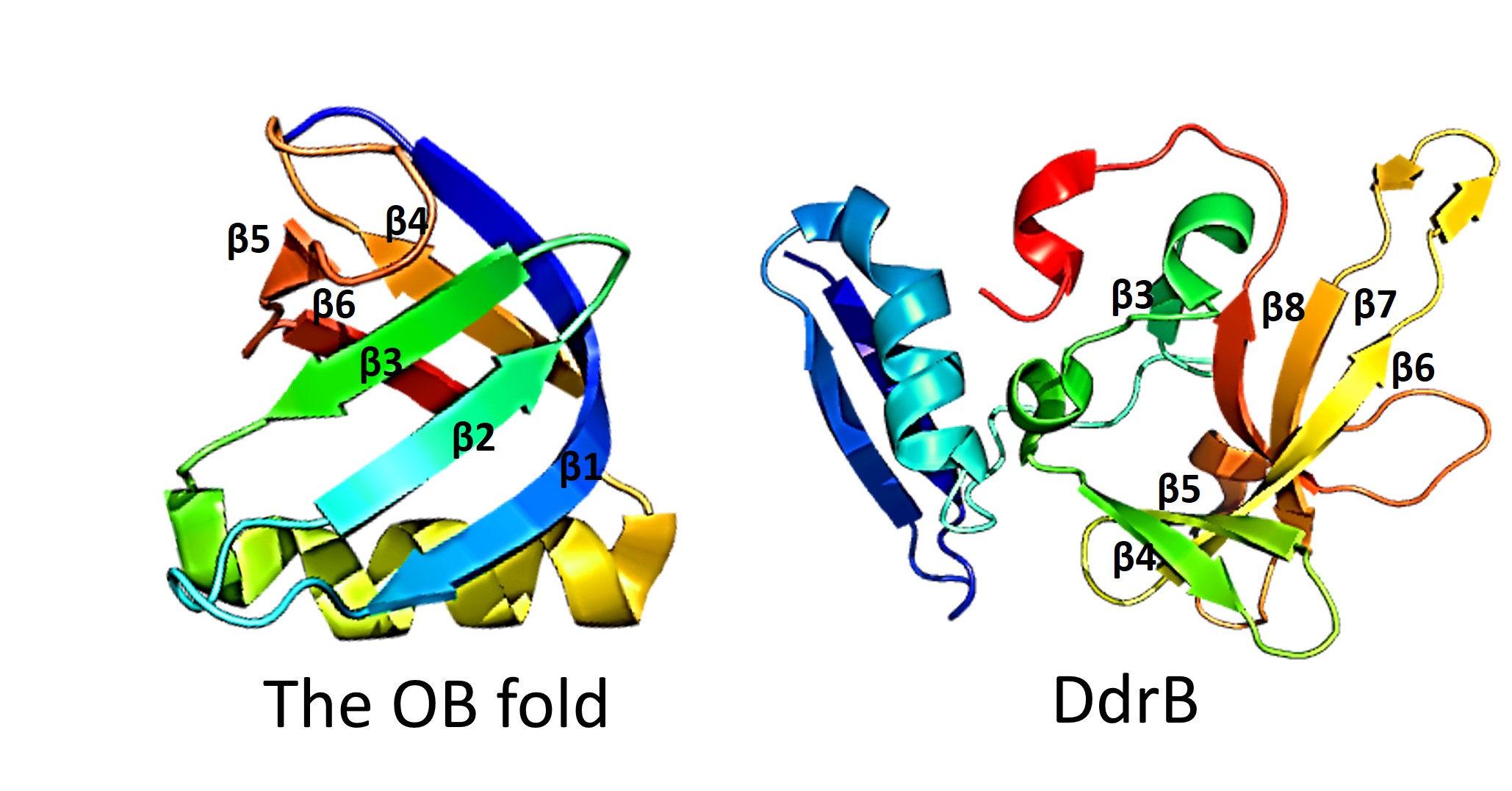
| 11:29, 30 April 2014 | OB fold.jpg (file) |  |
242 KB | This figure highlights the differences between the classic OB fold found in single-stranded binding proteins and the novel structural features of DdrB. The OB fold is observed in the protein verotoxin-1, PDB code 2XSC . DdrB is modeled from the PDB stru |
| 13:54, 28 April 2014 | Models.jpg (file) |  |
250 KB | From 4HQB. This was generated using pymol. |
| 13:39, 28 April 2014 | DNA channel.jpg (file) |  |
224 KB | From 4HQB. This was made using Pymol |
| 15:46, 18 April 2014 | 4HQB.pdb2.gz (file) |  |
411 KB | Test - not for use |
| 16:37, 1 April 2014 | 3f9f.pdb (file) | |
423 KB | |
| 15:46, 15 May 2012 | Intermediate.jpg (file) |  |
33 KB | Activated protein C tetrahedral intermediate |
| 15:40, 15 May 2012 | FV cleavage.jpg (file) |  |
45 KB | Forms of Factor V and Cleavage of Factor Va by activated protein C |
| 14:35, 15 May 2012 | Structral Homology.jpg (file) |  |
61 KB | Structural Homology with Vitamin K dependent Blood Coagulation Proteins |