Uploads by Mayank Mamgaain
From Proteopedia
Jump to navigationJump to search
This special page shows all uploaded files.
| Date | Name | Thumbnail | Size | Description |
|---|---|---|---|---|
| 09:32, 9 September 2020 | Iub2359-sup-0002-figures2.jpg (file) |  |
2.79 MB | Superimposition of the Cryo‐EM structure of SCV2‐nsp12:RNA and the model of the ternary complex. The backbone of the Cryo‐EM structure is colored green and that of the model is colored cyan. The RNA is shown in the stick representation. The incorpor |
| 09:29, 9 September 2020 | Iub2359-sup-0001-figures1.jpg (file) |  |
2.31 MB | Model of the functional ternary complex of the SCV2‐nsp12 enzyme in the complex. The RNA substrate and incoming GTP are shown in stick representation and colored according to element. The N‐terminal extension region and the palm, fingers, and thumb do |
| 09:02, 9 September 2020 | Figure 5.jpg (file) |  |
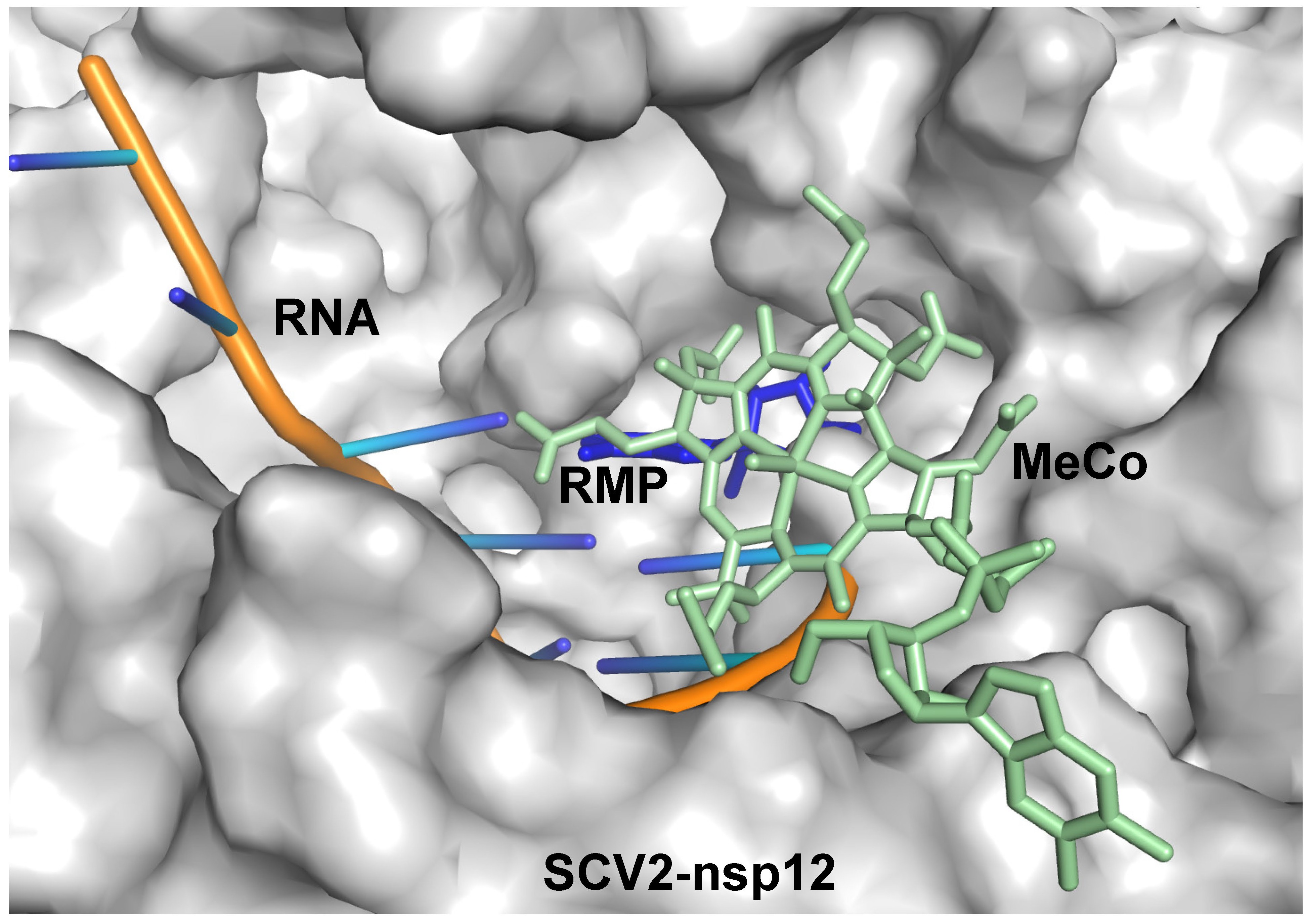
955 KB | Binding site of methylcobalamin (MeCo) overlaps with that of incoming nucleotide. The Cryo‐EM structure of SCV2‐nsp12 in complex with RNA bearing remdesivir monophosphate (RMP) at the 3′ end of the primer (7BV2) and the model of the SCV2‐nsp12:met |
| 07:54, 9 September 2020 | Figure 4B.jpg (file) |  |
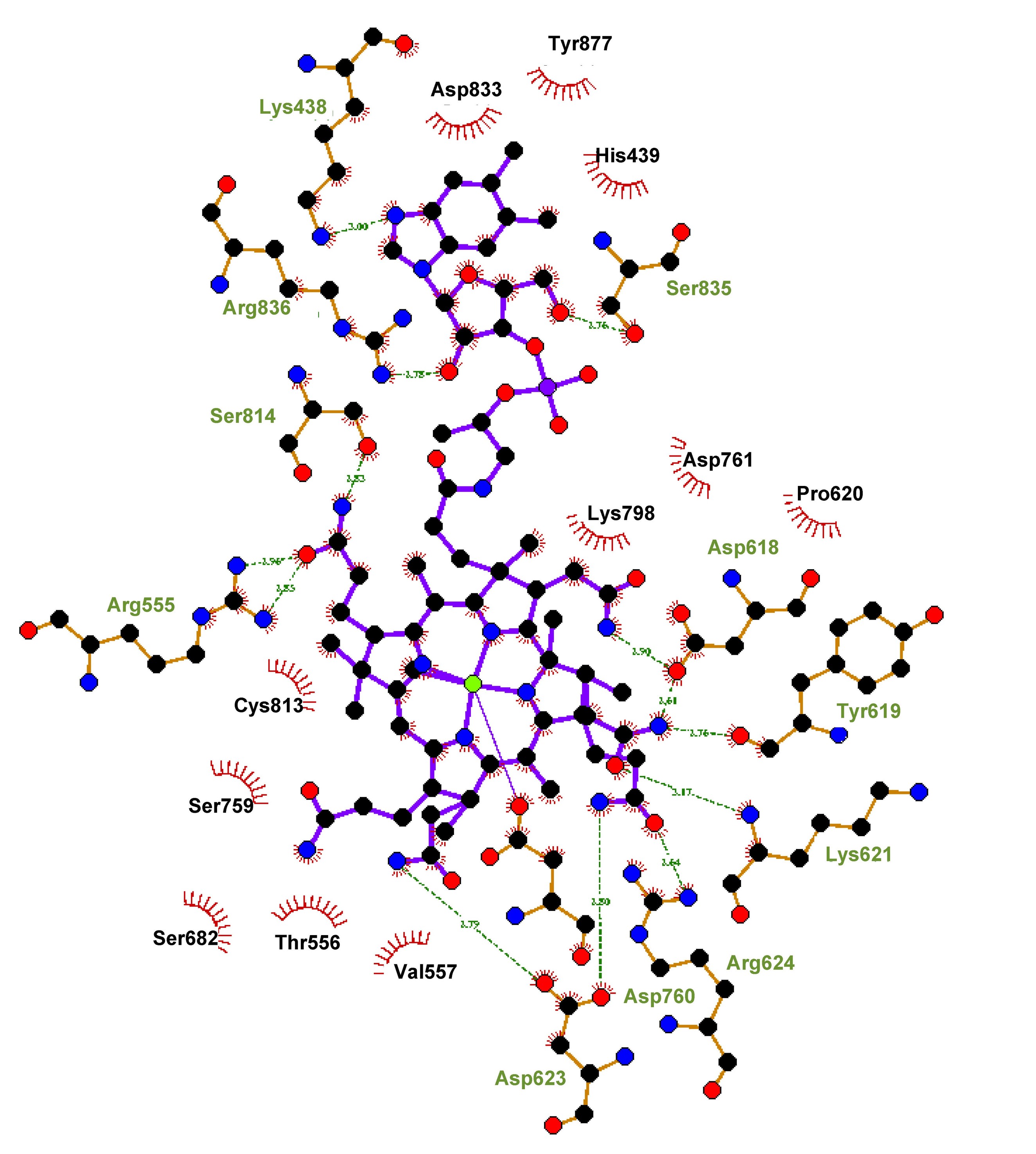
597 KB | Residues of SCV2‐nsp12 that interact with methylcobalamin. The LigPlot representation of the interacting residues is displayed here. The polar interactions are shown in the form of dotted lines and the residues involved in hydrophobic interactions are h |
| 07:22, 9 September 2020 | Figure 4A.jpg (file) |  |
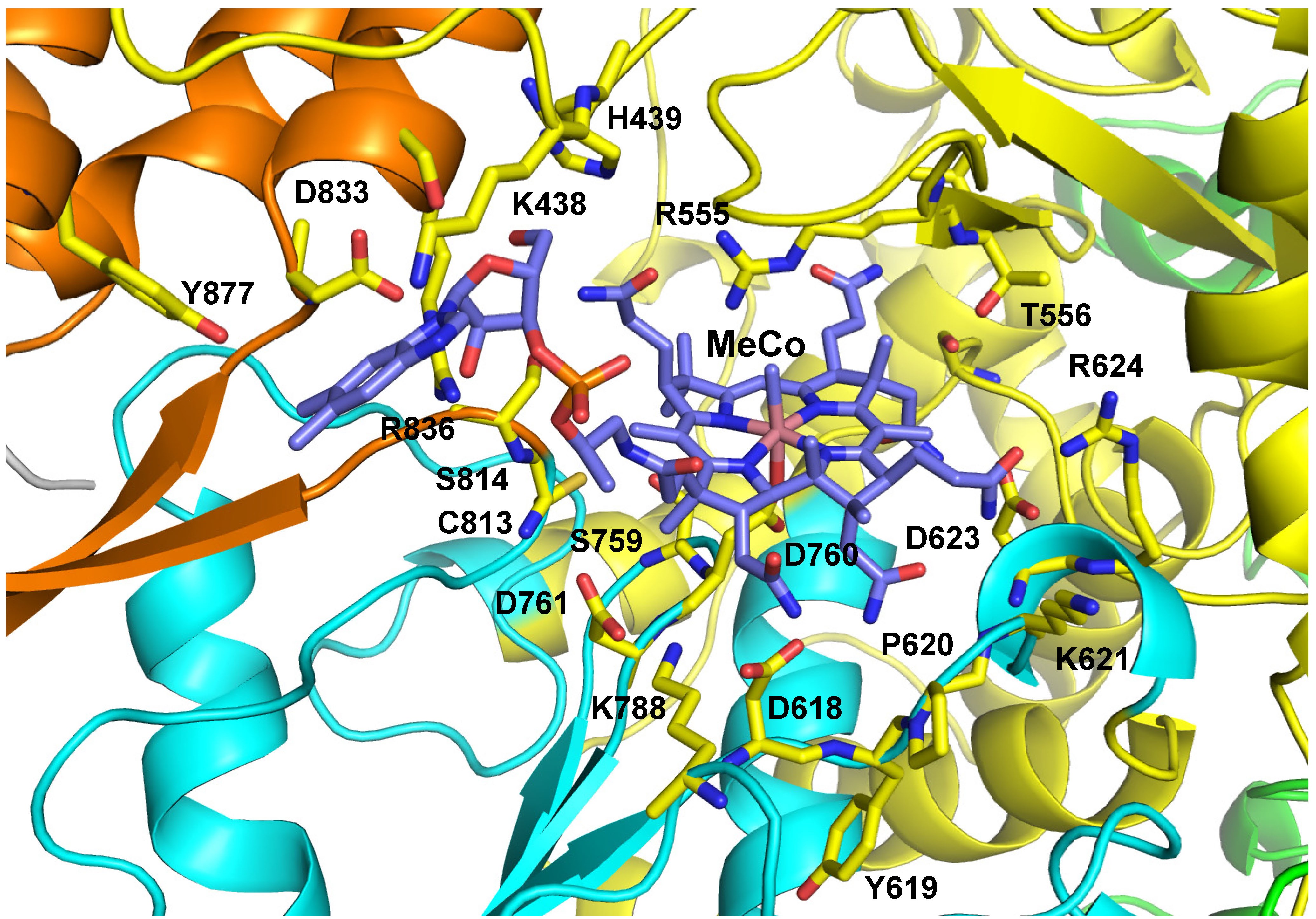
1.63 MB | Residues of SCV2‐nsp12 that interact with methylcobalamin. The vitamin B12 (MeCo) molecule is shown in stick representation and colored according to element. The N‐terminal extension region and the palm, fingers, and thumb domains are shown in green, |
| 06:28, 9 September 2020 | Figure 1a.jpg (file) |  |
883 KB | |
| 06:25, 9 September 2020 | Figure 1.jpg (file) |  |
883 KB | |
| 06:23, 9 September 2020 | SCV2‐nsp12.jpg (file) |  |
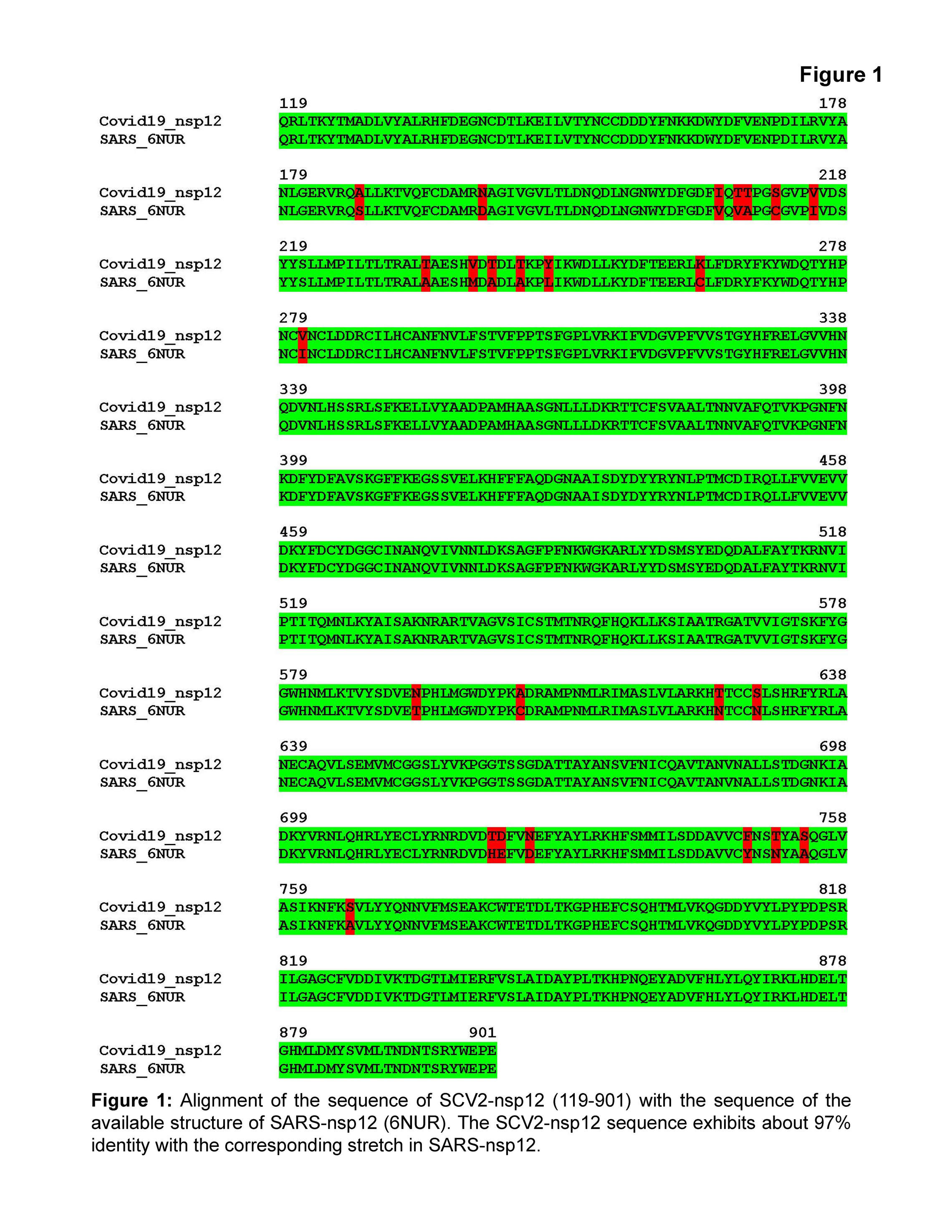
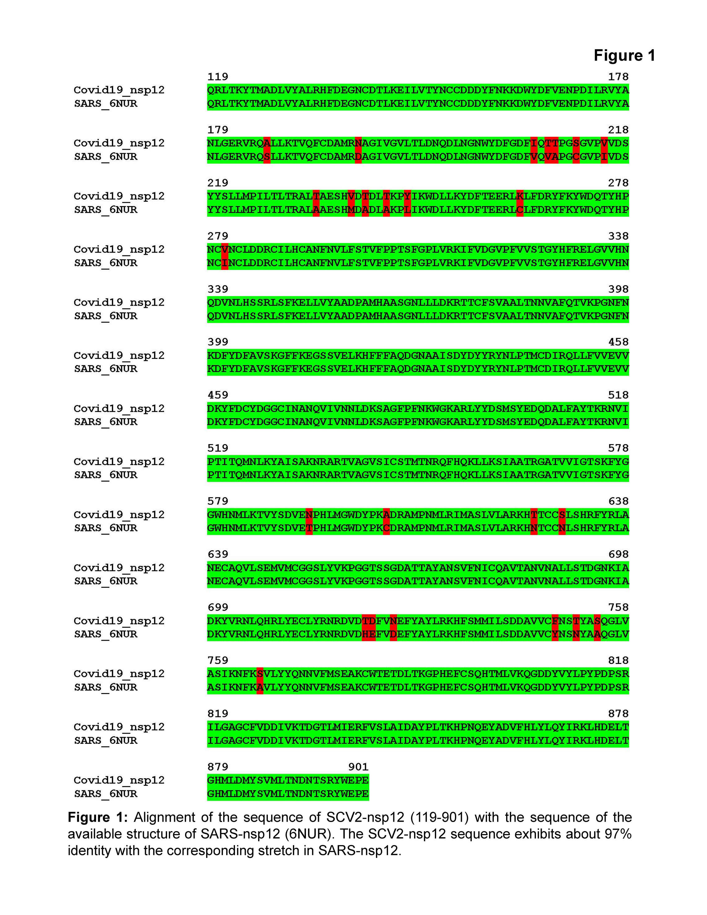
883 KB | Alignment of the sequence of SCV2‐nsp12 (119–901) with the sequence of the available structure of SARS‐nsp12 (6NUR). The SCV2‐nsp12 sequence exhibits about 97% identity with the corresponding stretch in SARS‐nsp12 |
| 06:13, 9 September 2020 | Figure 3.jpg (file) |  |
588 KB | Model of the SCV2‐nsp12 enzyme in complex with methylcobalamin. The vitamin B12 molecule is shown in stick representation and colored according to element. The N‐terminal extension region and the palm, fingers, and thumb domains are shown in green, cy |
| 09:35, 7 September 2020 | DeepakTNair PAC2020 Figure3.jpg (file) |  |
1.38 MB | Model of the SCV2‐nsp12 enzyme in its apostate. The N‐terminal extension region and the palm, fingers, and thumb domains are shown in green, cyan, yellow, and orange, respectively. The catalytic residues Asp618, Asp760, and Asp761 are colored accordin |
| 09:15, 7 September 2020 | Iub2359-fig-0001-m.jpg (file) |  |
1.51 MB |