Caspase-3/Sandbox
From Proteopedia
| |||||||||
| Crystal Structure of Unliganded Human Caspase-3 | |||||||||
|---|---|---|---|---|---|---|---|---|---|
| Gene: | CASP3 OR CPP32 (Homo sapiens) | ||||||||
| |||||||||
| |||||||||
| Resources: | FirstGlance, OCA, PDBsum, RCSB | ||||||||
| Coordinates: | save as pdb, mmCIF, xml | ||||||||
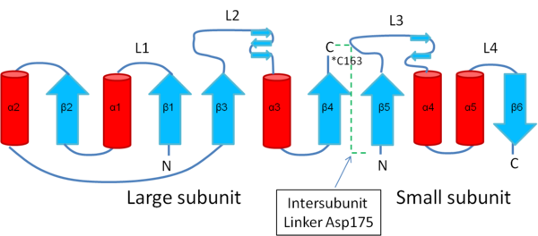
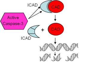
Caspases are proteases that function via a cysteine residue in the active site to cleave substrates after aspartic acid residues. Caspases are crucial for the initiation (e.g. caspase-8, -9, -10) and execution (e.g. caspase-3, -6, -7) of apoptosis, or programmed cell death either via the intrinsic or extrinsic pathway (Degterev, Boyce et al. 2003). In its inactive form, procaspase-3 consist of a large subunit and small subunit, interjected by an aspartic acid residue. This aspartate is the site recognized by activated initiator caspases, such as caspase-8 and caspase-9. Upon cleavage, procaspase-3 is separated into two subunits, p17/20 and p12/10 respectively, which heterodimerize to yield the active form of caspase-3. In its active form, caspase-3 is able to cleave substrates such as ICAD (inhibitor of caspase-activated deoxyribonuclease). Cleavage of ICAD leads to abrogation of its inhibitory effect on CAD, allowing CAD to migrate into the nucleus and cause double-strand breaks in DNA, thus contributing to apoptosis(Enari, Sakahira et al. 1998; Sakahira, Enari et al. 1998).
Contents |
Structure & Function
Structure
Procaspase-3, unlike initiator procaspases, is a stable dimer with little enzymatic activity. Procaspase-3 contains a tri-aspartate ‘safety-catch’ (Asp179–Asp181) in the intersubunit linker to remain nonfunctional. During the process of activation, this linker undergoes a pH-dependent conformational change, exposing Asp175 for cleavage. This leads to the formation of a heterotetramer (Walters, 2011). The heterotetramer consists of two anti-parallel arranged heterodimers, each one formed by a 17 kDa (p17) and a 12 kDa (p12) subunit (http://www.uniprot.org/uniprot/P42574).
Caspase-3 consists of a twisted, mostly parallel beta-sheet sandwiched between two layers of alpha-helices. The chains are classified as alpha/beta, with one chain containing a 3-layer(aba) sandwich with Rossmann fold topology and another chain containing a 2-layer sandwich with alpha-beta plaits(Fuentes-Prior and Salvesen, 2004).
Salt Bridge
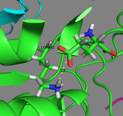
Lys242 is located at the C-terminus of helix 5 and Glu246 is located at the base of loop L4 of caspase-3. It has been suggested that the salt bridge between Lys242-Glu246 (NH3+ of Lys with COO- of Glu) is not found in the zymogen form of caspase-3 but forms only upon caspase-3 maturation. Rather, these residues are involved in other interactions in procaspase-3. Lys242 maintains the native procaspase dimer, whereas glutamate is involved in stabilizing loop L3, the substrate binding loop. In procaspase-3, Glu246 participates in hydrogen bonding with Trp214. In the mature, active caspase-3, the stability of the active site of caspase-3 depends on the salt bridge between Lys242 and Glu246 to maintain the correct conformation of loop L4. In caspase-3, Glu246 neutralizes the positive charge of Lys242 by forming a salt bridge within the same hydrophobic cluster of residues between helices 4 and 5 that buries the aliphatic portion of Lys242. Mutations of either K242A or E246A resulted in the complete loss of procaspase-3 activity as measured by fluorescence emission following addition of substrate (Ac-DEVD-AFC) and excitation at 400nm. These mutants were active in the mature, fully cleaved caspase-3, although to a much lower extent than wild type mature caspase-3, perhaps due to altered accessibility of its active site. The measurements of kcat/Km for these mutants at the optimal pH (7.5) for caspase-3 activity are 14 to 60-fold lower than that of wild type, with an overall increase in Km and a decrease in kcat. This may be due to alterations caused by the mutation(s) in the loop bundle interaction between loops L2, L4 and L2’. Loss of the Lys242-Glu246 salt bridge may alter the position of helix 4, which consequently alter the position of loop L3, leading to altered hydrogen-bonding in the P4 site. This may explain the decrease in activity seen in the mutants. Furthermore, because Phe250 in loop L4 participates in hydrogen bonding to P4, mutations in loop L4 could result in disruptions of these interactions (Feeney, 2004; Walters, 2011).
Posttranslational Modifications
Caspase-3 is translated as a zymogen. Activation occurs via cleavage by initiator caspases, such as caspase-8, -9, and -10, and by granzyme B to generate the p12(small) and p17(large) subunits. (http://www.uniprot.org/uniprot/P42574)
S-Nitrosylation
An ambient level of nitric oxide has antiapoptotic effects via S-nitrosylation of caspase-3 zymogens at its catalytic Cys163 residue perhaps because it directly associates with all 3 NOS isoforms. S-nitrosylation effectively inhibits the enzyme activity of caspase-3 (Mannick, 2007). NO and S-nitrosoglutathione (GSNO) reacts nonspecifically to all cysteine residues of caspase-3 (Mitchell, 2005). Caspase-3 nitrosylation at a second cysteine residue (Cys47, Cys220, or Cys 264) may have further antiapoptotic effects by leading to its association with acid sphingomyelinase (ASM). ASM inhibits cleavage and activation of caspase-3 by initiator caspases, such as caspase-8 and caspase-9 (Mannick, 2007).
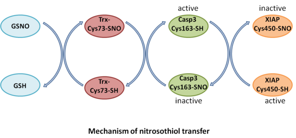
Thioredoxin, the main intracellular oxidoreductase, does not inhibit caspase-3 activity. However, Trx-Cys73-SNO inhibits apoptosis by decreasing caspase-3 activity via transnitrosylation at Cys163 catalytic residue. This reaction is reversible, although this is not as likely since Cys163 on caspase-3 is more nucleophilic than Cys73 on Trx. GSNO is capable of transferring its nitroso-group to Trx-Cys73, which in turn can S-nitrosylate caspase-3, leading to inactive caspase-3 (Mitchell, 2005).
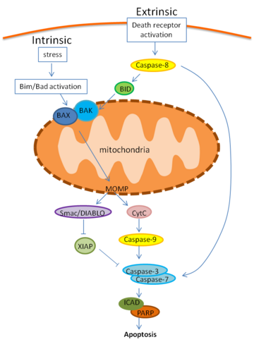
In human B- and T-cell lines, nitrosylation of caspase-3 depends on localization: mitochondrial but not cytoplasmic caspase-3 zymogens are S-nitrosylated. S-nitrosylation may prevent improper autoactivation in the mitochondria due to close proximity in the intermembrane space (Mannick, 2001). Mitochondrial caspase-3 is released into the cytoplasm and becomes denitrosylated when FasL binds Fas receptor (Mannick, 2001). Fas receptor activation by FasL activates the extrinsic pathway of apoptosis through activation of caspase-8, which can either directly cleave procaspase-3 to caspase-3 or lead to Bid cleavage to tBid. tBID then permeates into the mitochondria, leading to cytochrome c release, apoptosome formation with caspase-9, and finally caspase-3 cleavage to its activated form. Smac/DIABLO is also released from the mitochondria and leads to inhibition of XIAP (X-linked inhibitor of apoptosis), abrogating its inhibitory effect on caspase-3 activity within the cytosol. XIAP also functions as an E3 ubiquitin ligase, targeting caspase-3 for proteasomal degradation (Nakamura, 2010). Aside from this, Fas-induced apoptosis also leads to denitrosylation of caspase-3 and therefore activation by freeing its catalytic Cys residue (Mannick, 1999). Perhaps, this denitrosylation occurs via transnitrosylation of XIAP RING domain at Cys450, which in effect inhibits its E3 ligase activity. This further prolongs caspase-3 activity by blocking its degradation, and thus promotes increased cell death (Nakamura, 2010). Thus, S-nitrosylation of caspases inhibits caspase activation and apoptosis, whereas denitrosylation activates caspases and promotes apoptosis (Mannick, 2007).
Other
The first residue of caspase-3, Met1, is posttranslationally modified as N-acetylmethionine. Ser26 is also possibly modified as Phosphoserine.(http://www.uniprot.org/uniprot/P42574)
Active Site/Ligand Binding
| |||||||||||
Function
Caspases are proteases that function via a cysteine residue in the active site to cleave substrates after aspartic acid residues. Caspases are crucial for the initiation (e.g. caspase-8, -9, -10) and execution (e.g. caspase-3, -6, -7) of apoptosis, or programmed cell death either via the intrinsic or extrinsic pathway (Degterev, Boyce et al. 2003).
In the intrinsic pathway, stimuli such as trophic factor withdrawal, UV irradiation, chemotherapeutics, DNA damage, endoplasmic reticulum (ER) stress, activates B cell lymphoma 2 (BCL-2) homology 3 (BH3)-only proteins like Bim or Bad, leading to BCL-2-associated X protein (BAX) and BCL-2 antagonist or killer (BAK) activation and mitochondrial outer membrane permeabilization (MOMP). Following MOMP, cytochrome c is released and binds apoptotic protease-activating factor 1 (APAF1), inducing recruitment of the apoptosome and activation of caspase-9, an initiator caspase. Caspase-9 cleaves and activates effector caspases, caspase-3 and caspase-7, leading to apoptosis through cleavage of death substrates such as Inhibitor of Caspase-activated Deoxyribonuclease (ICAD) and Poly ADP ribose polymerase (PARP). Mitochondrial release of second mitochondria-derived activator of caspase (Smac/DIABLO) relieves the caspase inhibitory function of X-linked inhibitor of apoptosis protein (XIAP).
The extrinsic apoptotic pathway is initiated by the activation of death receptors, such as Fas by FasL. Fas-associated death domain protein (FADD) is recruited to the receptor along with caspase-8. This results in the dimerization and activation of caspase-8, which can then directly cleave and activate caspase-3 and caspase-7, leading to apoptosis. Crosstalk between the extrinsic and intrinsic pathways occurs through BH3-only protein BH3-interacting domain death agonist (BID) cleavage to truncated BID (tBID) by active caspase-8 (Li, Zhu et al. 1998). It has become appreciated more recently that caspase activity, specifically caspase-8, is also required for T cell growth (Alam, Cohen et al. 1999; Kennedy, Kataoka et al. 1999; Misra, Jelley-Gibbs et al. 2005), and that the location and level of active caspases within cells may be a key determinant of survival or death (Misra, Russell et al. 2007; Koenig, Russell et al. 2008). Murine αβ T cells bearing high levels of caspase activity manifest increased rates of both cell growth and cell death (Dohrman, Russell et al. 2005).
Caspase-3
In its inactive form, procaspase-3 consist of a large subunit and small subunit, interjected by an aspartic acid residue. This aspartate is the site recognized by activated initiator caspases, such as caspase-8 and caspase-9. Upon cleavage, procaspase-3 is separated into two subunits, p17/20 and p12/10 respectively, which heterodimerize to yield the active form of caspase-3. In its active form, caspase-3 is able to cleave itself as well as other caspase. It also cleaves substrates such as ICAD (inhibitor of caspase-activated deoxyribonuclease). Cleavage of ICAD leads to abrogation of its inhibitory effect on CAD, allowing CAD to migrate into the nucleus and cause double-strand breaks in DNA, thus contributing to apoptosis(Enari, Sakahira et al. 1998; Sakahira, Enari et al. 1998). Cleavage of poly(ADP-ribose) polymerase (PARP) between Asp216-Gly217 also contributes to apoptosis. (http://www.phosphosite.org/proteinAction.do?id=4672&showAllSites=true)
Disease
It has been suggested that caspases play a part in chronic (Alzheimer's, Huntington's, and Parkinson's diseases) and acute neurodegenerative disease. Inhibition of caspase activity increases survival of neurons in nerve crush injuries. Studies have also shown that caspases are downregulated to allow for unchecked survival of neoplastic cancers and autoimmune diseases. In particular, caspase-8 has been found to be silenced in neuroblastoma and humans with mutations in caspase-8 and -10 develop ALPS (autoimmune lymphoproliferative syndrome). These raise the possibility that caspase-3 may play a role as well. Knocking out caspase-3 in 129x1/SvJ mice leads to hydrocephalus and subsequently perinatal lethality. Caspase-3 C57BL/6J knockout mice however appear to have no brain abnormalities (Fuentes-Prior and Salvesen, 2004). SNO-caspases have also been suggested to promote cell injury and death in neurodegenerative disorders by transnitrosylating XIAP (Nakamura, 2010).
Evolutionarily Related Proteins
To date, eighteen caspases have been identified. Caspases can largely be grouped into three subfamilies in humans: inflammatory (caspase-1, -4, and -5), effector (caspase-3, -6, and -7), and initiator caspases (-2, -8, -9, and -10). Caspase-11 and -12 substitutes for caspase-4 and -5, respectively, in mice. (Fuentes-Prior and Salvesen, 2004)
Solved Structures
References & Notes
Alam, A., L. Y. Cohen, et al. (1999). "Early activation of caspases during T lymphocyte stimulation results in selective substrate cleavage in nonapoptotic cells." J Exp Med 190(12): 1879-90.
Aouad, S. M., L. Y. Cohen, et al. (2004). "Caspase-3 is a component of Fas death-inducing signaling complex in lipid rafts and its activity is required for complete caspase-8 activation during Fas-mediated cell death." J Immunol 172(4): 2316-23.
Budd, R. C. (2001). "Activation-induced cell death." Curr Opin Immunol 13(3): 356-62.
Degterev, A., M. Boyce, et al. (2003). "A decade of caspases." Oncogene 22(53): 8543-67.
Dohrman, A., J. Q. Russell, et al. (2005). "Cellular FLIP long form augments caspase activity and death of T cells through heterodimerization with and activation of caspase-8." J Immunol 175(1): 311-8.
Enari, M., H. Sakahira, et al. (1998). "A caspase-activated DNase that degrades DNA during apoptosis, and its inhibitor ICAD." Nature 391(6662): 43-50.
Feeney B, Pop C, Tripathy A, Clark AC. Ionic interactions near the loop L4 are important for maintaining the active-site environment and the dimer stability of (pro)caspase 3. Biochem J. Dec 15 2004;384(Pt 3):515-525.
Fuentes-Prior, P. and G. S. Salvesen (2004). "The protein structures that shape caspase activity, specificity, activation and inhibition." Biochem J 384(Pt 2): 201-232.
Kennedy, N. J., T. Kataoka, et al. (1999). "Caspase activation is required for T cell proliferation." J Exp Med 190(12): 1891-6.
Koenig, A., J. Q. Russell, et al. (2008). "Spatial differences in active caspase-8 defines its role in T-cell activation versus cell death." Cell Death Differ 15(11): 1701-11.
Li, H., H. Zhu, et al. (1998). "Cleavage of BID by caspase 8 mediates the mitochondrial damage in the Fas pathway of apoptosis." Cell 94(4): 491-501.
Mannick JB, Hausladen A, Liu L, et al. Fas-induced caspase denitrosylation. Science. Apr 23 1999;284(5414):651-654.
Mannick JB, Schonhoff C, Papeta N, et al. S-Nitrosylation of mitochondrial caspases. J Cell Biol. Sep 17 2001;154(6):1111-1116.
Mannick JB. Regulation of apoptosis by protein S-nitrosylation. Amino Acids. 2007;32(4):523-526.
Mitchell DA, Marletta MA. Thioredoxin catalyzes the S-nitrosation of the caspase-3 active site cysteine. Nat Chem Biol. Aug 2005;1(3):154-158.
Nakamura T, Wang L, Wong CC, et al. Transnitrosylation of XIAP regulates caspase-dependent neuronal cell death. Mol Cell. Jul 30 2010;39(2):184-195.
Misra, R. S., D. M. Jelley-Gibbs, et al. (2005). "Effector CD4+ T cells generate intermediate caspase activity and cleavage of caspase-8 substrates." J Immunol 174(7): 3999-4009.
Misra, R. S., J. Q. Russell, et al. (2007). "Caspase-8 and c-FLIPL associate in lipid rafts with NF-kappaB adaptors during T cell activation." J Biol Chem 282(27): 19365-74.
Sakahira, H., M. Enari, et al. (1998). "Cleavage of CAD inhibitor in CAD activation and DNA degradation during apoptosis." Nature 391(6662): 96-9.
Vincent, M. S., K. Roessner, et al. (1996). "Apoptosis of Fashigh CD4+ synovial T cells by borrelia-reactive Fas-ligand(high) gamma delta T cells in Lyme arthritis." J Exp Med 184(6): 2109-17.
Walters J, Swartz P, Mattos C, Clark AC. Thermodynamic, enzymatic and structural effects of removing a salt bridge at the base of loop 4 in (pro)caspase-3. Arch Biochem Biophys. Apr 1 2011;508(1):31-38.
Wang Z, Watt W, et al. (2010) Kinetic and structural characterization of caspase-3 and caspase-8 inhibition by a novel class of irreversible inhibitors. Biochim Biophys Acta 1804(9):1817-31.
Wesselborg, S., O. Janssen, et al. (1993). "Induction of activation-driven death (apoptosis) in activated but not resting peripheral blood T cells." J Immunol 150(10): 4338-45.

