Image:PPC Motif in SARS-CoV-2 Spike Protein.jpg
From Proteopedia

Size of this preview: 800 × 538 pixels
Full resolution (1280 × 861 pixel, file size: 130 KB, MIME type: image/jpeg)
Summary
PNAS May 26, 2020 117 (21) 11727-11734; first published May 6, 2020 https://doi.org/10.1073/pnas.2003138117
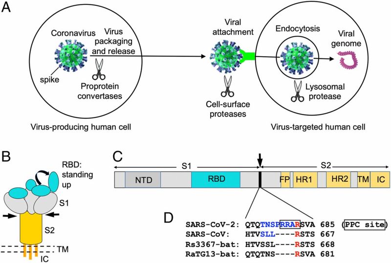
Figure 1. PPC motif in SARS-CoV-2 spike protein. (A) Different stages of coronavirus entry where host cellular proteases may activate coronavirus spikes. (B) Schematic drawing of the three-dimensional (3D) structure of coronavirus spike. S1, receptor-binding subunit; S2, membrane fusion subunit; TM, transmembrane anchor; IC, intracellular tail. (C) Schematic drawing of the 1D structure of coronavirus spike. NTD, N-terminal domain. FP (fusion peptide), HR1 (heptad repeat 1), and HR2 (heptad repeat 2) are structural units in coronavirus S2 that function in membrane fusion. (D) Sequence comparison of the spike proteins from SARS-CoV-2, SARS-CoV, and two bat SARS-like coronaviruses in a region at the S1/S2 boundary. Only SARS-CoV-2 spike contains a putative PPC motif—RRAR (residues in the box). The assumed PPC cleavage site is in front of the arginine residue labeled in red. The spike region mutated from SARS-CoV-2 sequence (TNSPRRA) to SARS-CoV sequence (SLL) is labeled in blue. GenBank accession numbers are QHD43416.1 for SARS-CoV-2 spike, AFR58740.1 for SARS-CoV spike, MG916901.1 for bat Rs3367 spike, and QHR63300.2 for bat RaTG13 spike.
Licensing
Creative Commons Attribution 3.0 License
![]()
File history
Click on a date/time to view the file as it appeared at that time.
| Date/Time | User | Dimensions | File size | Comment | |
|---|---|---|---|---|---|
| (current) | 19:41, 17 September 2020 | Jeremiah C Hagler (Talk | contribs) | 1280×861 | 130 KB | PNAS May 26, 2020 117 (21) 11727-11734; first published May 6, 2020 https://doi.org/10.1073/pnas.2003138117 Figure 1. PPC motif in SARS-CoV-2 spike protein. (A) Different stages of coronavirus entry where host cellular proteases may activate coronaviru |
- Edit this file using an external application
See the setup instructions for more information.
Links
There are no pages that link to this file.
