Image:PPC Motif in SARS-CoV-2 Spike Protein Cropped to Spike Schematic Only B.jpg
From Proteopedia
No higher resolution available.
PPC_Motif_in_SARS-CoV-2_Spike_Protein_Cropped_to_Spike_Schematic_Only_B.jpg (316 × 333 pixel, file size: 46 KB, MIME type: image/jpeg)
Summary
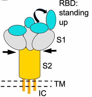
Shang et al., PNAS May 26, 2020 117 (21) 11727-11734; first published May 6, 2020 https://doi.org/10.1073/pnas.2003138117
Figure 1B Only: Schematic drawing of the three-dimensional (3D) structure of SARS-CoV-2 coronavirus spike. S1, receptor-binding subunit; S2, membrane fusion subunit; TM, transmembrane anchor; IC, intracellular tail.
Licensing
Creative Commons Attribution 3.0 License
![]()
File history
Click on a date/time to view the file as it appeared at that time.
| Date/Time | User | Dimensions | File size | Comment | |
|---|---|---|---|---|---|
| (current) | 19:54, 17 September 2020 | Jeremiah C Hagler (Talk | contribs) | 316×333 | 46 KB | PNAS May 26, 2020 117 (21) 11727-11734; first published May 6, 2020 https://doi.org/10.1073/pnas.2003138117 Figure 1B Only: Schematic drawing of the three-dimensional (3D) structure of SARS-CoV-2 coronavirus spike. S1, receptor-binding subunit; S2, mem |
- Edit this file using an external application
See the setup instructions for more information.
Links
The following pages link to this file:
