Image:Pdbnmr.jpg
From Proteopedia
Size of this preview: 800 × 450 pixels
Full resolution (1152 × 648 pixel, file size: 50 KB, MIME type: image/jpeg)
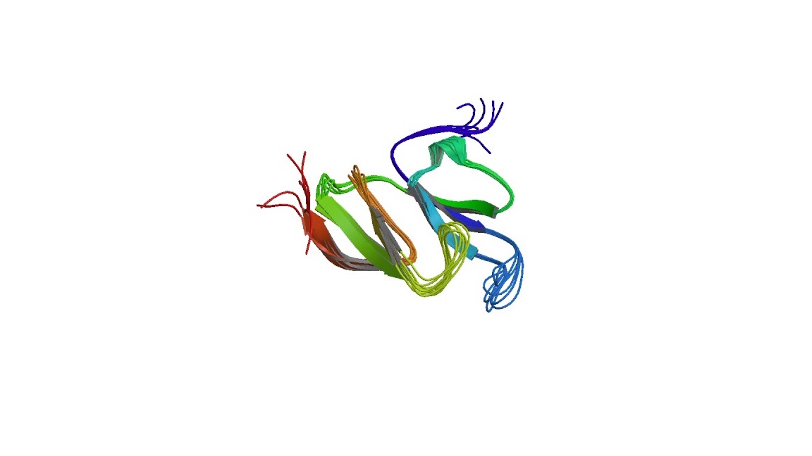
The structure of the two domain fragment of MVP, depicted by NMR. Taken from the 1Y7X entry in the PBD. credit to: Kozlov, G., Vavelyuk, O., Minailiuc, O., Banville, D., Gehring, K., and Ekiel, I. (2006) Solution structure of a two-repeat fragment of major vault protein. J. Mol. Biol. 356, 444 – 452.
File history
Click on a date/time to view the file as it appeared at that time.
| Date/Time | User | Dimensions | File size | Comment | |
|---|---|---|---|---|---|
| (current) | 06:49, 17 March 2018 | Idan Ben-Nachum (Talk | contribs) | 1152×648 | 50 KB | The structure of the two domain fragment of MVP, depicted by NMR. Taken from the 1Y7X entry in the PBD. credit to: Kozlov, G., Vavelyuk, O., Minailiuc, O., Banville, D., Gehring, K., and Ekiel, I. (2006) Solution structure of a two-repeat fragment of majo |
- Edit this file using an external application
See the setup instructions for more information.
Links
The following pages link to this file:
