Transpeptidase (TP), also known as penicillin-binding proteins (PBP), catalyze the cross-linking of peptidoglycan polymers during bacterial cell wall synthesis. Beta-lactam (β-lactam) antibiotics, which
include penicillins, cephalosporins and carbapenems, bind and irreversibly inhibit transpeptidases. The overuse and misuse of β-lactam antibiotics has led to strains of Staphylococcus aureus that are resistant to all β-lactams and are often only susceptible to “last resort antibiotics”, such as vancomycin.
Cell Wall Structure
The cell wall, which is composed of peptidoglycan, is crucial for maintaining the structural integrity of the bacterium. Peptidoglycan consists of N-acetylmuramic Acid (NAM) and N-acetylglucosamine (NAG) polymers. Rows of peptidoglycan are cross-linked together with pentaglycine chains. The NAM residues have a five amino acid side chain that terminates with two D-Alanine (D-Ala) residues.
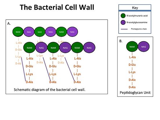
Figure 1. A.Bacterial Cell Wall B.Peptidoglycan with D-Ala-D-Ala substrate
Structure of a Resistant Transpeptidase
Methicillin resistant Staphylococcus aureus (MRSA) is resistant to all β-lactams because it acquires an alternative PBP, PBP2a, that is not bound or inhibited by any β-lactams. PBP2a is composed of two domains: a non-penicillin binding domain and a transpeptidase binding domain . The NBP domain of PBP2a is anchored in the cell membrane, while the TP domain “sits” in the periplasm with its active site facing the inner surface of the cell wall. The active site contains which catalyzes the cross-linking of the peptidoglycan rows with pentaglycine cross-links.
Catalytic Mechanism of PBP2a
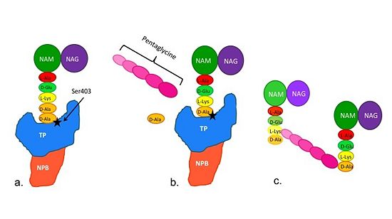
Figure 2. Schematic diagram illustrating the mechanism of action of PBP2a
(a)The D-Ala-D-Ala side-chain substrate of the peptidoglycan accesses the active site of the PBP2a.
(b)Ser403 nucleophilically attacks the peptide bond of the terminal D-Ala residues of the substrate. The terminal D-Ala residue then exits the active site. The now terminal D-Ala residue forms a covalent bond to Ser403, while a crosslinking pentaglycine chain enters the active site.
(c)A covalent bond forms between the pentaglycine chain and the terminal D-Ala residue, regenerating the active site serine residue.
The entire process takes 4 milliseconds.
How Do Antibiotics Work?
The β-lactam antibiotics inhibit bacterial growth by inhibiting PBPs and ultimately cell wall
synthesis. Specifically, β-lactams are molecular mimics of D-Ala-D-Ala, which is the normal
substrate of PBPs. Nucleophillic attack of the β-lactam results in the PBP being irreversibly
inhibited by the β-lactam. As a result, the synthesis of the cell wall is inhibited which leads
to cell lysis.
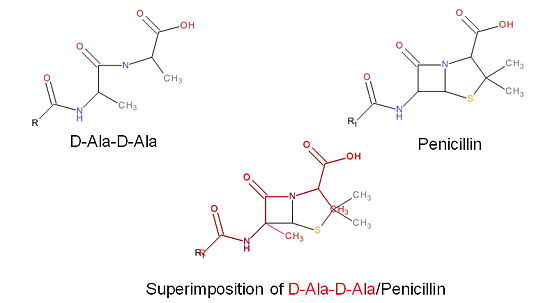
Figure 3. Mechanism of action of β-lactams. A. Structure of a β-lactam (penicillin) showing the amide, carboxyl, and β-lactam ring groups β-lactam ring groups. B. Structure of the D-Ala-D-Ala substrate. C. Overlay of the D-Ala-D-Ala substrate in red with penicillin demonstrating molecular mimicry.
PBP2a and Ceftobiprole
MRSA becomes resistant to β-lactams by acquiring an alternative PBP, PBP2a, that is
neither bound nor inhibited by β-lactams. Recently, two cephalosporins – and ceftaroline – that have anti-MRSA activity have been developed. Ceftobiprole is able to
inhibit PBP2a because additional chemical groups at the position of the cephalosporin backbone are able to interact with additional amino acid residues in PBP2a; specifically
. As a result of its tighter binding to PBP2a, ceftobiprole is able to more
efficiently react with the serine active site residue and therefore inhibit the activity of
PBP2a.
PBP2a and Ceftaroline
(PDB:3ZFZ)
In addition to TP domain of PBP2a, there is an allosteric domain,highlighted orange, in which the distance between is 60Å.
Allosteric site serves as a binding site for the substrate . When the substrate binds to the (Tyr105, Asn146, Asp295, Tyr297), a conformational change occurs at the active site, opening it and allowing catalytic action to occur.
The medicine, , mimics the substrate at the allosteric site opening the active site, allowing to .


