Anaplastic Lymphoma Kinase (ALK)
Introduction

Figure 1. Outline of the domains and regions of anaplastic lymphoma kinase
Anaplastic lymphoma kinase is a receptor tyrosine kinase (RTK) important to the regulation of functions within the central nervous system [1]. RTKs are the high-affinity cell surface receptors for many polypeptide growth factors, cytokines, and hormones. ALK activates pathways that promote cell growth and proliferation, similar to the Insulin Receptor (IR). These pathways include, but are not limited to, the ERK, JAK, and PI3K pathways. ALK is composed of two identical monomers consisting of seven unique domains, and two intermixed regions. Among these, one of the most notable is the glycine-rich region (GlyR), characterized by helical structures formed by glycine residues. Other domains are archetypal of RTK activation and regulation. Several potential human ALK ligands have been discovered, however two ALK activating ligands (ALKALs) have been further studied for their ability to activate ALK, ALK homologs, and ALK fusion proteins [2]. The preferred ALKAL for activation of the kinase domain is AUG, which binds in a dimeric fashion to ALK, inducing a conformational change. In order to determine the atomic details of human ALK dimerization and activation by AUG, the methods of cryo-electron microscopy, nuclear magnetic resonance, and X-ray crystallography were utilized [1]. Due to its role in cell growth and proliferation signaling, ALK is considered to be a proto-oncogene, meaning that it is capable of producing mutations that lead to cancer. ALK mutations can lead to various types of cancers, including non-small-cell lung cancer, anaplastic large cell lymphoma, squamous cell carcinoma, and inflammatory myofibroblastic cancer [3].
General Structure

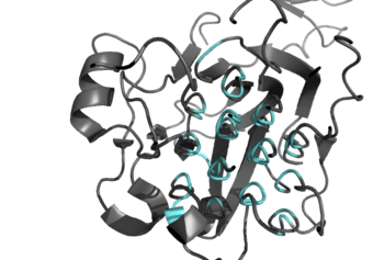
Figure 2: Shown in teal is the Glycine Rich Region of ALK in its helical structure.
Anaplastic lymphoma kinase is a , in which each contains seven domains and two regions [4]. These domains and regions are as follows: N-terminal region (NTR), two meprin–A-5 protein–receptor protein tyrosine phosphatase μ domains (MAM), low density lipoprotein receptor class A domain (LDL), (TNF), (GlyR), (EGF), transmembrane α-helix (TMH), kinase domain [1]. The NTR functions as a signal peptide, the structure of which is yet to be determined. Both MAM domains and the LDL domain are unique to ALK within the superfamily of RTKs, and the biological role of these domains has not yet been determined [2]. However, the LDL and MAM regions are thought to be involved in various regulatory functions; studies of other LDL protein domains indicate a role for LDL domains in plasma cholesterol homeostasis [5]. Additionally, research on MAM domains implicates a role in cell-cell interactions through homophilic binding [2]. The TNF-like domain assists in mediating mature T-cell receptor induced apoptosis. Both the TNF domain and GlyR region are discontinuous, traversing each other frequently [1]. The GlyR region consists of several glycine helices, a highly unique structure. Though the function of ALK's EGF domain is unknown, EGF domains are typically found in the extracellular region and are thought to be important building blocks for extracellular proteins [6]. The TMH connects the extracellular and intracellular regions of ALK through the plasma membrane. The kinase domain is in the intracellular region and is autoactivated through phosphorylation at positions . This allows the RTK to begin signaling cascades throughout the cell [6]. While the structures of the N-terminal region, MAM, and LDL have not been determined, it has been shown that only the TNF, GlyR, and EGF portions of ALK are required in the extracellular region for ligand activation via ALKALs [3]. All portions of anaplastic lymphoma kinase are located in the extracellular domain except for the transmembrane α-helix, which is in the transmembrane region, and the kinase domain located in the intracellular region.
Ligand Binding

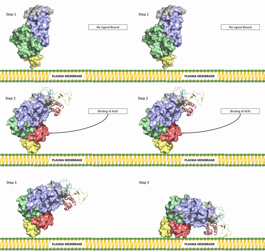
Figure 3: Gif-image of the conformational change occurring in the extracellular region of Anaplastic Lymphoma Kinase once the AUG ligand has bound to the ligand-binding site. This change is stabilized through contacts of the AUG and the plasma membrane. The video was made using stop motion animation techniques, then converted to gif format using EZgif.
ALKALs recognized by ALK are FAM150, binding in either a monomeric or dimeric fashion, and in a dimeric fashion [7] . Its biologically preferred ligand is AUG, a protein ligand containing 128 residues that form three α-helices. FAM150 is a structural ortholog of AUG. The binding of ALK to it's ligand results in homodimerization and an induced conformational change. Prior to the ligand binding to ALK, the extracellular domain is oriented vertically and perpendicularly to the plasma membrane (Step 1, Figure 3). Once the ligand is (Step 2, Figure 3), ALK undergoes a conformational change and folds over so that the on the ALKAL can now interact with the negatively charged phosphate heads of the plasma membrane (Step 3, Figure 3). The residues of ALK and its ligand interact through the formation of [8]. It has been hypothesized that the GlyR region plays a role in the flexibility needed to complete the conformational change. This conformational change via ligand activation allows for the auto-activation of the kinase domain, where the domains use the tyrosine phosphorylation mechanism to phosphorylate tyrosine residues on the opposite monomer. ALK is a Class II RTK (Insulin Receptor (IR) Family), sharing much of its kinase activation mechanism with the IR.
Tyrosine Phosphorylation Mechanism

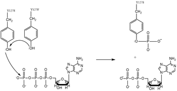
Figure 4. Tyrosine phosphorylation mechanism
Dimerization of anaplastic lymphoma kinase activates the of each monomer [7]. Next, the kinase domains phosphorylate the tyrosine residues (Y1278, Y1282, and Y1283 [9]) of the opposite monomer using the hydrolysis of ATP into ADP and Pi [8]. These phosphorylated tyrosine residues recruit signal proteins via phosphorylation mechanisms similar to that of the IR. These signal proteins start signaling cascades through various signal pathways including the ERK, JAK, and PI3K pathways. These pathways signal for cell proliferation and survival (ex: begin transcription). The mechanism of tyrosine phosphorylation is a key step in the signal activation, transduction, and regulation of ALK enzymatic activity. This mechanism is similar to Class II RTKs, used by the IR in insulin signal activation and transduction. Mutations in catalytic tyrosine residues leads to non-functional ALK [2]. In the absence of a bound ligand, it is proposed that the kinase domain of ALK is instead cleaved by Caspase 3 (Casp 3,) which subsequently induces apoptosis [3].
Applications

Figure 5. Structure of the ALK inhibitor, Crizotinib
Characterization of ALK as a proto-oncogene implicated in various types of cancers has made it an attractive target for drug therapies [10]. Treatments are designed to correct overactivation of the kinase domain of ALK, one of the main observations in ALK-positive cancers. ALK-positive cancers are those in which the individual has an oncogenic mutation in the ALK protein sequence that contributes to the proliferation of cancer cells. Overactivation of the kinase domain is caused by aberrant ATP binding, leading to the upregulation of cell growth signaling pathways. This overexpression of growth signals allows cancerous cells to grow and divide more rapidly than normal cells. One therapeutic medication seeing increased use with high levels of success is . Approved by the FDA in January of 2021, Crizotinib treats pediatric/young adult ALK-positive anaplastic large cell lymphoma. The treatment has an overall response rate of 90%, working by to the ATP binding site of the kinase domain [11]. Crizotinib preferentially binds to ATP in this region, therefore effectively blocking the binding of ATP to the kinase domain [11]. This prevents the signal transduction via intercellular signaling pathways, which works to prevent cancerous cells from growing and spreading. Other possible inhibition mechanisms of Crizotinib suggest the inhibition of the RTK, cMET, as the ATP binding site of both kinase domains being very similar both sequentially and structurally [12]. Apart from possible inhibition of cMET, Crizotinib is regarded as a highly successful in regard to its specificity for the ATP binding site of the ALK kinase domain [12].


