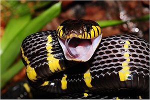
Figure 1. Mangrove catsnake
![Figure 2. Three fingers formed by three peptide loops (F1, F2 & F3) [PDB=2H5F]](/wiki/images/thumb/3/3f/Kolme_sormea_figure_text.png/300px-Kolme_sormea_figure_text.png)
Figure 2. Three fingers formed by three peptide loops (F1, F2 & F3) [PDB=2H5F]
Denmotoxin is a snake venom protein produced by Boiga dendrophila (mangrove catsnake) which belongs to a family of well studied three-fingered neurotoxins.
Denmotoxin is a member of three-finger toxin family
Three-finger toxins (3FTXs) are the most common family of snake venom proteins; these venoms can be found in elapid, colubrid and hydrophiid snakes and include toxins such as α-cobratoxin and α-bungarotoxin. 3FTXs are non-enzymatic proteins which form a structurally conserved superfamily whose members all share a highly conserved structure. [1][2] The core structure of 3FTXs is formed by three joined together by four conserved disulphide bridges located in the of the protein. Despite the similarities in the structure of different toxins belonging to the family, the 3FTXs from various venoms have a variety of receptors/acceptors and exhibit differential responses in their targets. The members of the family can vary slightly in: the length and type of twists of the tree loops; the length and type of turns of the N and C-terminal tail and the amount of β-sheets in the overall structure. These differences allow for the specificity and toxicity of the proteins to their targets. [3]
Denmotoxin shares approximately 30% sequence similarity with other 3FTXs with an exception of exhibiting approximately 50% sequence similarity with another colubrid snake venom α-colubritoxin. Despite the relatively low sequence similarity, denmotoxin possesses all the residues needed to maintain the 3 finger fold. A large part of the sequence similarity between denmotoxin and other 3FTXs is due to the highly conserved disulphides and a number of structurally important residues. Denmotoxin belongs to a subdivision of non-conventional three-finger toxins; the novel features of denmotoxin are discussed below. [3]
Structure of the non-conventional 3FTX denmotoxin
Denmotoxin is a monomeric protein comprising of 77 amino acid residues. Denmotoxin consists of (fig. 2) protruding from the ; this structure is typical for 3FTXs. The globular core consists of a triple stranded anti-parallel β-sheet; two of the β-strands in this structure connect to the second loop () and one β-strand connects to the third loop. There are two highly on the protein: one at the tip of the central loop and one at the 3 first residues of the N-terminus; the expected of denmotoxin is at the tip of the . [3]
There are 10 structurally important cysteine-residues in denmotoxin which form five stabilizing . Four of these disulphide bonds, which are found in all 3FTXs are located at the central core and the is found at the tip of the first loop. The cysteine residues of all 3FTXs are highly conserved, whereas the other residues within the sequence express high variability. Denmotoxin possesses most of the conserved residues invariant among 3FTXs which have been shown to be important for the proper folding of and structure of protein. The presence of these structurally important residues result in the characteristic three finger fold of the toxin (e.g. Gly52, Pro58). [3]
Multiple sequence alignment of denmotoxin reveals that the venom belongs to the family of non-conventional 3FTXs. All non-conventional 3FTXs have an additional disulphide bond. Denmotoxin has 7 additional amino acid residues in its N-terminal when compared to other 3FTXs; the N-terminus is also blocked by a pyroglutamic acid residue. This unusually long N-terminus is unstructured and is hypothesized to gyrate above the core of the protein. Another unique feature of denmotoxin is the twist at the tip of the central loop originating from a kink in a (Pro40). At the central loop, the charge is also negative; an arginine residue has been replaced with an aspartic acid, which is unusual for the proteins of the family. [3]
Interaction model with nicotinic acetylcholine receptor
![Figure 3. Suggested interaction of α-bungarotoxin in binding pocket of nAChR (simplified figure) [PDB=4HQP]](/wiki/images/thumb/d/d7/Bungarotoxin_interaction_with_nACHr_figure_text.png/300px-Bungarotoxin_interaction_with_nACHr_figure_text.png)
Figure 3. Suggested interaction of
α-bungarotoxin in binding pocket of nAChR (simplified figure) [PDB=4HQP]
Most of the 3FTX family proteins possess a highly conserved arginine in the tip of their binding loop responsible for interaction with nAChRs. This interaction was determined by resolving a structure of α7 nAChR chimera in a complex with α-bungarotoxin (fig. 3) [4]. In denmotoxin, this loop contains several changes in its sequence; arginine has replaced by aspartic acid (ASP41) and two additional glutamates (GLU42 & GLU45) generate untypical into the loop. [3]
Biochemistry of denmotoxin is unique for its taxon specificity to bird nicotinic acetylcholine receptors (nAChR). Binding of denmotoxin to chick muscle AChR (α1βγδ) is a highly irreversible whereas interaction with identical subunit assembly in mouse AChR is reversible. The reversible binding allows the receptor to function properly, but in the case of irreversible binding nAChR is prevented of natural agonist activation. Previous studies [5] with 3FTXs have shown that the binding of toxin leads to “locking down” of the nACh receptor, preventing required conformational change for ion channel activation and induction of signal. [3]
There are no significant differences in the sequence of functionally important loops A-F of nAChR in mice and chicks. However in the prior region of loop F, chicks have several changes in their amino acid composition leading to introduction of positive charge spatially in the front of the functionally active loop F. This might have important functionality in the attraction and binding of denmotoxin specifically to bird nAChRs. However, no crystal structure of denmotoxin binding to nAChR has been resolved; thus denmotoxin could also utilise a yet uncharacterised binding site in the receptor. [3]
Conclusions
3FTXs are the most abundant and well characterized snake venom protein family. The broad spectrum of 3FTX targets, including nAChRs, phospholipids and L-type calcium channels, make the proteins interesting for research. [3] The interaction of 3FTX-proteins with their targets result in a myriad of pharmacological effects. Study of the 3FTX family proteins can also be utilised in the research of their receptors. Understanding how the architecture of the proteins of the family result in the varying mechanisms of action can prove a valuable tool for rational drug design.
Whilst the highly conserved three finger structural motif is present in denmotoxin minor structural differences can be found when compared to other proteins of the family. These differences are responsible for the recognition of target receptors and can modulate both the specificity and toxicity of the venom.

![Figure 2. Three fingers formed by three peptide loops (F1, F2 & F3) [PDB=2H5F]](/wiki/index.php/Image:Kolme_sormea_figure_text.png)
![Figure 3. Suggested interaction of α-bungarotoxin in binding pocket of nAChR (simplified figure) [PDB=4HQP]](/wiki/index.php/Image:Bungarotoxin_interaction_with_nACHr_figure_text.png)
