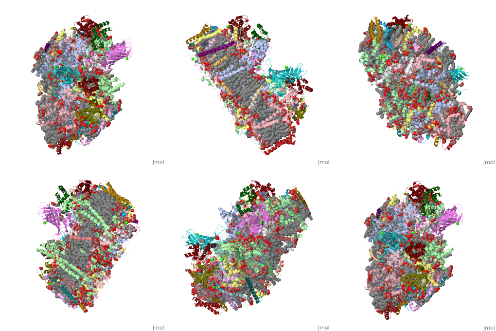
Structural highlights
3wu2 is a 20 chain structure with sequence from Thermostichus vulcanus. Full crystallographic information is available from OCA. For a guided tour on the structure components use FirstGlance.
|
| Method: | X-ray diffraction, Resolution 1.9Å |
| Ligands: | , , , , , , , , , , , , , , , , , , , , |
| Resources: | FirstGlance, OCA, PDBe, RCSB, PDBsum, ProSAT |
Function
PSBA_THEVL D1 (PsbA) and D2 (PsbD) bind P680, the primary electron donor of photosystem II (PSII) as well as electron acceptors. PSII is a light-driven water plastoquinone oxidoreductase, using light energy to abstract electrons from H(2)O, generating a proton gradient subsequently used for ATP formation.[HAMAP-Rule:MF_01379]
Publication Abstract from PubMed
Photosystem II is the site of photosynthetic water oxidation and contains 20 subunits with a total molecular mass of 350 kDa. The structure of photosystem II has been reported at resolutions from 3.8 to 2.9 A. These resolutions have provided much information on the arrangement of protein subunits and cofactors but are insufficient to reveal the detailed structure of the catalytic centre of water splitting. Here we report the crystal structure of photosystem II at a resolution of 1.9 A. From our electron density map, we located all of the metal atoms of the Mn(4)CaO(5) cluster, together with all of their ligands. We found that five oxygen atoms served as oxo bridges linking the five metal atoms, and that four water molecules were bound to the Mn(4)CaO(5) cluster; some of them may therefore serve as substrates for dioxygen formation. We identified more than 1,300 water molecules in each photosystem II monomer. Some of them formed extensive hydrogen-bonding networks that may serve as channels for protons, water or oxygen molecules. The determination of the high-resolution structure of photosystem II will allow us to analyse and understand its functions in great detail.
Crystal structure of oxygen-evolving photosystem II at a resolution of 1.9 A.,Umena Y, Kawakami K, Shen JR, Kamiya N Nature. 2011 May 5;473(7345):55-60. Epub 2011 Apr 17. PMID:21499260[1]
From MEDLINE®/PubMed®, a database of the U.S. National Library of Medicine.
See Also
References
- ↑ Umena Y, Kawakami K, Shen JR, Kamiya N. Crystal structure of oxygen-evolving photosystem II at a resolution of 1.9 A. Nature. 2011 May 5;473(7345):55-60. Epub 2011 Apr 17. PMID:21499260 doi:10.1038/nature09913

