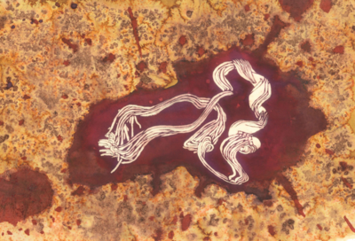
Art:Ebola GP fusion loop
From Proteopedia
Behind the Artwork and the Protein Structure
The artist who created this image is Anna Valchanova, a student at Stephen Perse School in Cambridge, UK. She has presented the protein inside a drop of blood to reflect the blood transmission of the disease, and depicted the protein structure in bleach to reflect the decline of health once infected with Ebola virus. Anna has based her work on the GP ‘fusion loop’ structure, solved by NMR spectroscopy in the Tamm lab, in the University of Virginia, School of Medicine.
View in 3D or go to PDB structure 2m5f [1]
Ebola Virus
The Ebola virus is a member of the family Filoviridae that form filamentous infectious virions, and encode their genome in single-stranded RNA. Ebola virus can be transmitted from wild animals to humans and also spreads through human-to-human transmission. To infect a host cell, the membrane of the virus fuses with that of the host, then the contents of the virus - its genome and proteins - enter the cell. After infection and replication in the host cell, the virus’ emerge from the cell, the surface of the virus is then formed from a membrane which is taken from the host cell. While the membrane might contain many host cell proteins, the only Ebola protein on the surface of the virus is one known as glycoprotein, or GP.
The Glycoprotein of Ebola
GP is shaped like a three pronged bowl and is anchored in the viral membrane by three helices, and is the protein responsible for getting the virus into the cytoplasm of a host cell. Mature GP is composed of two chains, termed GP1 and GP2, linked by a disulphide bond, between two cysteine amino acids. GP1 is responsible for binding to host cell receptors during infection and forms most of the bowl while GP2 sits at the base of the bowl, tying the trimer together and mediating the fusion of the viral and host membranes. A crucial part of Ebola GP2 is the fusion loop. It lies flat against the surface of the GP trimer until a drop in pH trigers a conformational change. This change enables the tip of the loop to insert into host cell membranes and triggers the membrane fusion process.
Human antibodies, found in Ebola survivors (PDB entry 3csy), bind to the fusion loop These antibodies may act by preventing the change in shape of the loop and thus blocking the virus entering the human cells. The fusion loop in GP is also the site of action of drugs such as toremifene (PDB entry 5jq7) and ibuprofen (PDB entry 5jqb) which are thought to destabilise the fusion process and could lead to Ebola therapueutics.
Once the tip of the fusion loop has inserted into the host membrane, GP2, including the fusion loop, undergoes a huge change in conformation. The post-fusion conformation of GP2 (PDB entry 1ebo) is vastly different to that before membrane fusion and shows that the fusion loop becomes helical and moves to the vicinity of the viral membrane, likely dragging the host membrane with it and effecting membrane fusion.
Explore the scientific publication
- ↑ Gregory SM, Larsson P, Nelson EA, Kasson PM, White JM, Tamm LK. Ebolavirus Entry Requires a Compact Hydrophobic Fist at the Tip of the Fusion Loop. J Virol. 2014 Apr 2. PMID:24696482 doi:http://dx.doi.org/10.1128/JVI.00396-14

