Art:Zika virus
From Proteopedia
Behind the Artwork and the Protein Structure
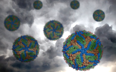
First isolated in the Zika Forest of Uganda in 1947, the image is that of Zika virus. The capsid, showing the beautiful icosahedral symmetry common to many viruses, is visually stunning, belying the disease it can cause. This image was created by Alice Clark at PDBe.
View in 3D or go to PDB structure 5ire [1]
A Zika outbreak in Brazil
Zika virus is related to other mosquito-borne viruses which cause dengue, yellow fever and West Nile fever. This group, called the Flaviviridae, have an RNA genome and are transmitted to humans via a mosquito vector, but Zika can also be transmitted between humans sexually. While Zika infection, if it causes symptoms at all, is usually mild, the virus has been linked to Guillain-Barre syndrome, an autoimmune disorder. There is growing evidence that in pregnant women it causes brain defects in their unborn babies.
Even though it has been known for 70 years, Zika came to prominence following the 2015 outbreak in Brazil leading to an epidemic in the Americas. Before this epidemic, there were no structures of Zika macromolecules in the PDB. The cryoEM structure of the virus capsid from the Rossmann group (PDB entry 5ire) published in Science in March 2016 was the first.
The Zika Proteome
The Zika genome encodes one long polyprotein which is 3400 amino acids long. After translation, this is cut up into the different functional portions, two of which (called E and M) form the capsid, and others, termed ‘non-structural proteins’ (hence the prefix NS) are necessary for the virus to replicate. Such has been the interest in Zika, that at the time of writing, less than a year after the first Zika structure arrived, there are now 45 different structures of Zika macromolecules in the PDB archive. More are added almost weekly; here’s the complete list. These structures cover much of the Zika proteome, and even a non-coding RNA (PDB entry 5tpy) from the 3’ untranslated region of its RNA genome.
They include structures of capsid proteins with antibodies bound, indicating which regions of the virus might be most useful in generating vaccines, and how antibodies stop the virus. There are also enzymes with small molecules bound, which could lead to future antivirals. Recently, a further cryoEM structure from the Rossmann group of immature virus (PDB entry 5u4w) has shown how the capsid is formed.
In two years since the disease came to prominence, scientists have structurally characterised much of Zika virus’ inner workings. Hopefully this puts the world in a strong position to find a vaccine and eradicate the virus.
Explore the scientific publication
- ↑ Sirohi D, Chen Z, Sun L, Klose T, Pierson TC, Rossmann MG, Kuhn RJ. The 3.8 A resolution cryo-EM structure of Zika virus. Science. 2016 Apr 22;352(6284):467-70. doi: 10.1126/science.aaf5316. Epub 2016, Mar 31. PMID:27033547 doi:http://dx.doi.org/10.1126/science.aaf5316

